
Evidence-based clinical review
Small Fiber Neuropathy: When EMG Is Normal but the Patient Is Not
A practical framework for recognizing the phenotype, confirming small-fiber dysfunction, finding treatable causes, and avoiding diagnostic overreach

Abstract
Background: Small fiber neuropathy (SFN) affects thinly myelinated A-delta fibers and unmyelinated C fibers that carry pain, temperature, and autonomic signals. Pure SFN can produce disabling sensory and autonomic symptoms while routine nerve conduction studies remain normal because they primarily assess larger myelinated fibers. [1,3]
Objective: This article provides a clinician-facing framework for deciding when a normal electromyography and nerve conduction study is compatible with SFN, how to obtain objective support, how to investigate causes without indiscriminate testing, and how to set evidence-based treatment limits. [1,2,7,10]
Key findings: A normal electrodiagnostic study neither invalidates the patient’s symptoms nor establishes SFN. Diagnostic confidence is strongest when a compatible phenotype and examination are paired with objective evidence from skin biopsy, quantitative sensory testing, autonomic testing, or another validated modality, with each test interpreted according to what it actually measures. [1,2,3,4,5] Skin biopsy is the best-validated direct structural test, but no single test is a stand-alone gold standard, and a biopsy does not identify the cause. [1,3,4,5] Etiologic assessment should be staged, prioritizing common metabolic, nutritional, toxic, paraproteinemic, immune, and infectious causes before phenotype-driven second-line testing. [7,8] Treatment should address an identified cause, neuropathic pain, autonomic dysfunction, function, and safety. [10,11] Randomized trials and AANEM guidance do not support routine intravenous immunoglobulin for idiopathic SFN or for SFN associated only with TS-HDS or FGFR3 antibodies. [12,13,14]
Conclusion: The practical goal is not to label every unexplained burning symptom as SFN. It is to recognize a plausible small-fiber phenotype, exclude a broader neurologic disorder, obtain objective support when it will change management, and pursue causes and treatments in proportion to the evidence. [1,2,6,10]
Introduction
The phrase “the EMG was normal” often becomes a diagnostic dead end. For a patient with burning feet, electric shocks, allodynia, altered sweating, orthostatic symptoms, or unexplained thermal sensory loss, that conclusion may be premature because routine electrodiagnostic studies do not directly test the small sensory and autonomic fibers implicated in SFN. [1,3]
The opposite error is equally important. Normal electrodiagnostic testing does not prove SFN, and symptoms alone do not establish the diagnosis. [1,2] The clinician must determine whether the pattern is anatomically plausible, whether examination findings support small-fiber dysfunction, whether another disorder better explains the presentation, and whether objective testing will change the next decision. [1,2,3,6]
What small fibers do
Small somatic fibers include thinly myelinated A-delta fibers and unmyelinated C fibers. They participate in pain and temperature sensation, while autonomic small fibers contribute to sudomotor, vasomotor, cardiovascular, gastrointestinal, genitourinary, and other visceral functions. [1,3,6]
A length-dependent SFN usually begins distally in the feet and may ascend over time, sometimes reaching the hands after substantial lower-extremity involvement. [6,8] Non-length-dependent SFN may be patchy, asymmetric, proximal, truncal, facial, or diffuse and should prompt closer attention to ganglionopathy, immune disease, infection, toxin exposure, or hereditary disease rather than automatically attributing it to a common distal polyneuropathy. [6,8,9]
Why EMG and nerve conduction studies can be normal
In everyday conversation, “EMG” often refers to the combined electrodiagnostic evaluation of nerve conduction studies and needle electromyography. Nerve conduction studies record responses from large myelinated sensory and motor fibers, while needle EMG evaluates motor-unit and muscle electrical activity. [1,3]
Pure SFN can therefore coexist with normal sensory and motor nerve-conduction results and a normal needle examination. [1,3] Electrodiagnostic testing remains useful because it can reveal a mixed large-fiber neuropathy, entrapment neuropathy, radiculopathy, plexopathy, motor neuropathy, or myopathy that changes both diagnosis and treatment. [1,3,10]
A normal study should be read as “no electrodiagnostic evidence of the large-fiber or motor abnormalities tested,” not as “no neurologic disease” and not as “SFN confirmed.” [1,3]
Recognizing a credible small-fiber phenotype
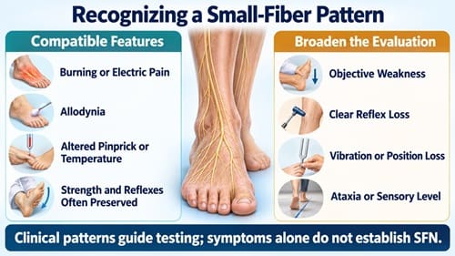
Common sensory complaints include burning, prickling, pins-and-needles sensations, electric or stabbing pain, painful cold, contact allodynia, hyperalgesia, and reduced pain or temperature perception. [1,6] Autonomic complaints may include altered sweating, color or temperature changes, orthostatic intolerance, palpitations, gastrointestinal dysmotility, urinary or sexual dysfunction, and pupillary symptoms. However, each of these has a broad differential diagnosis. [1,6]
Examination in pure SFN may show reduced pinprick or temperature sensation, hyperalgesia, allodynia, or autonomic skin changes. Strength, deep tendon reflexes, vibration, proprioception, and gait are often relatively preserved because these functions depend more heavily on motor or large sensory fibers. [1,2,3]
The following findings should broaden the evaluation beyond pure SFN: objective weakness, clear reflex loss, substantial vibration or joint-position loss, sensory ataxia, an extensor plantar response, cranial neuropathy, marked asymmetry, a sensory level, sphincter dysfunction suggesting myelopathy or cauda equina disease, or an abnormal nerve conduction study. [1,3,6]
| Pattern | Supports a pure SFN hypothesis | Suggests a broader or alternative process |
| Symptoms | Burning, electric pain, thermal dysesthesia, allodynia, autonomic complaints | Fixed dermatomal pain, focal entrapment pattern, prominent weakness, sensory level, severe ataxia |
| Examination | Pinprick or temperature abnormality with preserved strength, reflexes, vibration, and proprioception | Weakness, reflex loss, marked vibration or position loss, upper motor-neuron signs, cerebellar signs |
| Electrodiagnostics | Normal results can be compatible with pure SFN | Abnormal sensory or motor responses indicate large-fiber or mixed involvement. |
| Tempo | Chronic length-dependent progression is common | Acute or subacute onset, rapid progression, asymmetry, or systemic illness requires accelerated evaluation. |
The table is a clinical sorting aid, not a substitute for localization or a formal diagnostic standard. [1,2,3,9]
Objective confirmation: choose the test that answers the question
Skin biopsy
A 3-mm punch skin biopsy, usually obtained at the distal leg and sometimes at an additional proximal site, can quantify intraepidermal nerve fiber density (IENFD). Reduced IENFD below an appropriately validated lower reference limit supports structural small-fiber loss. [1,4,5]
Skin biopsy is the most established direct structural test for SFN, but interpretation depends on correct site selection, tissue processing, counting methods, and age-, sex-, and laboratory-appropriate normative data. [1,4,5] A reduced IENFD does not identify the cause, and a normal biopsy does not fully exclude early, patchy, non-length-dependent, or site-mismatched disease. [1,3,6]
Quantitative sensory testing
Quantitative sensory testing (QST) measures detection or pain thresholds for thermal and other stimuli. It can support small-fiber dysfunction, but it is psychophysical, requires attention and cooperation, and may be influenced by central nervous system processing, medications, mood, sleep, and testing technique. [2,3]
QST should therefore be interpreted as complementary functional evidence rather than a stand-alone locator of peripheral nerve pathology. [2,3]
Autonomic testing
Autonomic testing can be useful when the phenotype includes sweating abnormalities, orthostatic intolerance, or other objective autonomic concerns. Sudomotor testing, cardiovagal testing, and adrenergic testing examine different components of autonomic function and should be selected according to the symptom pattern and available laboratory expertise. [1,3,6]
An abnormal autonomic test supports dysfunction in the tested pathway, but it does not by itself prove that all symptoms arise from SFN or establish the etiology. [1,3]
Emerging and specialized techniques
Laser-evoked potentials, contact heat-evoked potentials, corneal confocal microscopy, microneurography, and other techniques may provide valuable information in specialized centers or research settings. Their availability, standardization, reference ranges, and reimbursement remain variable, so they should not be treated as interchangeable substitutes for a complete clinical assessment. [3,6]
A practical diagnostic-confidence framework
No single classification is universally mandated across laboratories and specialties. This framework is intended to clarify clinical reasoning, not to create new formal criteria. [1,2,3]
| Diagnostic level | Typical evidence | Clinical implication |
| Clinically suspected SFN | Compatible symptoms plus plausible distribution, with no better explanation; examination may be normal or show small-fiber signs | Review mimics and decide whether objective testing will change management |
| Objectively supported SFN | Compatible phenotype plus abnormal IENFD, QST, autonomic testing, or another validated small-fiber measure | Proceed with staged etiologic assessment and targeted treatment |
| Mixed neuropathy or alternate localization | Large-fiber signs, abnormal NCS/EMG, focal entrapment, radiculopathy, myelopathy, or another explanatory disorder | Reclassify the problem and treat the broader diagnosis |
| SFN not established | Symptoms without compatible signs or objective support, or internally discordant testing | Avoid diagnostic closure; reassess medications and consider central pain, musculoskeletal, vascular, dermatologic, and other causes. |
A strong diagnosis usually rests on convergence: a plausible phenotype, examination findings when present, exclusion of major alternatives, and objective evidence from at least one appropriately selected test. [1,2,3]
Finding the cause without ordering every test
The workup should begin with the history rather than the laboratory order set. The clinician should review diabetes and glucose intolerance, weight and metabolic risk, alcohol use, bariatric or gastrointestinal disease, nutritional intake, renal and hepatic disease, thyroid disease, infection risk, autoimmune symptoms, cancer history, family history, and exposure to chemotherapy, neurotoxic drugs, excess vitamin B6, heavy metals, or other toxins. [7,8]
Medication and supplement reconciliation deserves special attention. Over-the-counter products, energy drinks, compounded preparations, and multiple vitamin products can produce clinically important cumulative exposures that may not appear on the prescription list. [7,8]
A staged laboratory approach reduces both missed treatable causes and false-positive cascades. Adapt the panel to the phenotype, local prevalence, previous results, comorbidities, and pretest probability. [7,8]
| Stage | Commonly considered tests | When to expand |
| Initial metabolic and nutritional assessment | CBC; comprehensive metabolic panel; fasting glucose and HbA1c; vitamin B12 with methylmalonic acid when B12 status is uncertain; TSH | Consider an oral glucose tolerance test when dysglycemia remains suspected despite nondiagnostic routine testing; check vitamin B6 when supplements or exposure raise concern. |
| Paraprotein and immune assessment | Serum protein electrophoresis with immunofixation; additional monoclonal-protein testing when indicated; ANA and extractable nuclear antigen testing when systemic features are present; Sjögren and celiac testing when clinically compatible | Expand to complement, cryoglobulins, ANCA, inflammatory markers, or disease-specific testing when the phenotype or examination supports it. |
| Infectious assessment | HIV and hepatitis C testing according to risk, clinical context, consent, and local recommendations | Add other infectious tests only when exposure, geography, immune status, or systemic findings support them. |
| Phenotype-driven second tier | Amyloid evaluation, genetic testing, sarcoid evaluation, toxin testing, paraneoplastic assessment, or tissue/organ-directed studies | Early onset, family history, cardiomyopathy, nephropathy, bilateral carpal tunnel syndrome, unexplained weight loss, rapidly progressive autonomic failure, cancer clues, or other red flags |
This is not a universal panel. Broad autoantibody, infectious, genetic, or paraneoplastic testing in a low-pretest-probability patient can generate results that are difficult to interpret and may lead to unnecessary treatment. [7,8]
An abnormal risk factor should not automatically be declared causal. Mild dysglycemia, a low-titer antibody, or a monoclonal protein may be relevant, incidental, or one contributor among several; causality is more credible when the timing, phenotype, severity, and disease-specific evidence align. [7,8]
Red flags that change the pace of evaluation
Acute or subacute onset, rapid progression, marked asymmetry, non-length-dependent spread, prominent autonomic failure, systemic inflammatory symptoms, constitutional symptoms, objective weakness, ataxia, cranial nerve involvement, or new bowel and bladder dysfunction should prompt expedited reassessment rather than routine follow-up. [6,8,9]
Acute-onset SFN has been described, but observational evidence does not establish a single immune mechanism or justify automatic immunotherapy. [9] The immediate task is to confirm localization and look for infection, toxin exposure, nutritional disturbance, inflammatory disease, vasculitic or infiltrative neuropathy, spinal or root disease, and other time-sensitive alternatives. [7,8,9]
Treatment: cause-directed care first
When a credible cause is found, treatment should target it. Examples include improving diabetes management, stopping a neurotoxic exposure, correcting a documented nutritional deficiency without creating another toxicity, treating an infection, and applying disease-specific therapy for a confirmed systemic autoimmune, amyloid, genetic, renal, hepatic, or endocrine disorder. [7,8,10]
Cause-directed treatment may prevent further injury, but symptom resolution is not guaranteed because small-fiber damage may be only partly reversible and multiple mechanisms may coexist. [8,10]

Symptomatic treatment of neuropathic pain
Most medication evidence used in SFN is extrapolated from other neuropathic pain conditions, especially painful diabetic polyneuropathy. This distinction should be discussed explicitly because efficacy, labeling, and insurance coverage for another pain condition do not constitute approval for SFN as a syndrome. [10,11]
Reasonable first-line classes include tricyclic antidepressants, serotonin-norepinephrine reuptake inhibitors, gabapentinoids, and selected sodium-channel blockers. [11] Topical lidocaine or capsaicin may be useful when pain is localized, and systemic adverse effects are a concern, although product indications and the evidence base vary. [10,11]
Choice should be individualized according to kidney and liver function, orthostatic symptoms, cardiac disease, fall risk, anticholinergic burden, weight effects, sleep, mood, drug interactions, and patient priorities. [10,11] When an adequate trial produces no meaningful benefit or unacceptable adverse effects, switching to a different effective class is generally more rational than escalating an ineffective regimen indefinitely. [11]
Routine long-term opioid therapy should not be the default approach to chronic SFN pain. The AAN recommends against opioids for painful diabetic neuropathy, and the balance of limited durable benefit, tolerance, dependence, and overdose risk argues for similar caution when treating SFN-related pain. [11]
Nonpharmacologic and safety-focused care
Treatment should include function and safety, not only a pain score. Foot inspection, protection from heat and cold injury, appropriate footwear, fall-risk assessment, graded activity, physical therapy when deconditioning or gait impairment is present, sleep treatment, and attention to depression, anxiety, and pain-related coping can reduce disability even when nerve regeneration is uncertain. [10,11]
Patients with autonomic symptoms may need separate evaluation and treatment for orthostatic intolerance, sweating disorders, gastrointestinal dysfunction, or genitourinary symptoms. Therapy should be based on the documented autonomic disorder because many medications used for pain can worsen sedation, orthostasis, constipation, urinary retention, or falls. [1,6,10]
Immunotherapy: where the evidence draws a boundary
Empiric intravenous immunoglobulin should not be used routinely for painful idiopathic SFN. In a double-anonymized randomized trial of biopsy-confirmed idiopathic SFN, IVIG did not significantly improve pain or key secondary outcomes compared with placebo. [12]
A placebo-controlled pilot trial also did not detect benefit from IVIG in patients selected for TS-HDS or FGFR3 antibodies. [13] The updated AANEM consensus statement therefore advises against IVIG for idiopathic SFN and for SFN associated only with these antibodies. [14]
These findings do not mean that every neuropathy with small-fiber involvement is immune-treatment resistant. A patient with an established systemic autoimmune disease, vasculitic neuropathy, inflammatory ganglionopathy, or another defined immune neurologic disorder should be managed according to the disease-specific diagnosis and evidence, usually with neurology and the relevant specialty involved. [14,16]
Emerging reports of other immunotherapies remain preliminary. A 2025 rituximab series included only five antibody-positive cases and had no randomized control group, so it should be viewed as hypothesis-generating rather than a basis for routine treatment. [15]
Follow-up and referral
Follow-up should track pain, sleep, function, work or daily activity, autonomic symptoms, medication adverse effects, and objective neurologic change. [10,11] New weakness, reflex loss, gait ataxia, loss of vibration or proprioception, or abnormal nerve conduction findings suggest evolution into a mixed or alternative neuropathy and should trigger reassessment. [1,3,10]
Repeat skin biopsy or repeated broad laboratory panels are not routine outcome measures. They are most useful when a specific diagnostic or research question is likely to change management. [1,3,10]
Neurology or neuromuscular referral is appropriate when objective confirmation is needed, the phenotype is atypical, symptoms progress rapidly, large-fiber or motor findings appear, the cause remains unclear after a focused evaluation, or immunotherapy is being considered. [1,7,10] Autonomic-laboratory referral is appropriate when objective autonomic characterization will change treatment, while rheumatology, hematology, oncology, genetics, cardiology, nephrology, or an amyloid center may be needed for disease-specific red flags. [7,8,16]
Common diagnostic errors
- Using a normal EMG/NCS as proof that nothing neurologic is wrong. Pure SFN can be electrodiagnostically silent because the tested fibers are not the fibers primarily affected. [1,3]
- Using a normal EMG/NCS as proof of SFN. Normal testing excludes only the abnormalities that the study was designed and technically able to detect. [1,3]
- Diagnosing SFN from symptoms alone. Burning pain and dysautonomia are real but nonspecific; objective support and exclusion of mimics improve diagnostic reliability. [1,2]
- Treating skin biopsy as an etiologic test. Reduced IENFD demonstrates structural loss but does not distinguish diabetes, toxicity, autoimmunity, infection, hereditary disease, or idiopathic SFN. [1,4,5]
- Ordering every possible laboratory test at once. Low-pretest-probability testing increases incidental findings and can create false causal narratives. [7,8]
- Equating antibody positivity with a treatment indication. TS-HDS or FGFR3 antibodies alone have not established IVIG responsiveness in randomized evidence. [13,14]
- Escalating pain medication without measuring function or adverse effects. A regimen that does not produce meaningful benefit should be reconsidered, not merely intensified. [10,11]
A practical clinical algorithm
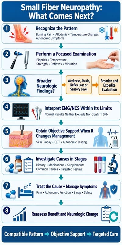
- Localize the syndrome. Decide whether symptoms fit a length-dependent, non-length-dependent, focal, autonomic-predominant, or mixed pattern. [1,6]
- Look for findings inconsistent with pure SFN. Weakness, reflex loss, proprioceptive loss, ataxia, upper motor-neuron signs, or a sensory level require a broader evaluation. [1,3]
- Use EMG/NCS for the question it can answer. Confirm or exclude large-fiber, motor, root, plexus, entrapment, or muscle involvement. [1,3,10]
- Obtain objective small-fiber support when it will change management. Choose skin biopsy, QST, autonomic testing, or a specialized modality based on phenotype and local expertise. [1,2,3,4,5]
- Investigate causes in stages. Start with history-guided common causes, then add targeted second-line tests for atypical features or red flags. [7,8,9]
- Treat the cause when established and manage symptoms safely. Use class-based neuropathic pain treatment, functional rehabilitation, and autonomic care while monitoring benefit and harm. [10,11]
- Reserve immunotherapy for a defined, evidence-supported immune diagnosis. Do not use idiopathic labeling or isolated TS-HDS/FGFR3 positivity as a substitute for that standard. [12,13,14,15,16]
Limitations of the evidence
SFN research remains limited by heterogeneous case definitions, referral-center populations, variable normative data, imperfect test agreement, and a shortage of disease-modifying randomized trials. [1,2,3,6] Diagnostic tests measure different domains, so discordant results are not unusual and should be resolved through clinical reasoning rather than by choosing whichever result confirms the preferred diagnosis. [1,2,3]
Treatment evidence for all-cause SFN is substantially thinner than the evidence for painful diabetic polyneuropathy. Clinicians therefore select many commonly used pain medications by extrapolation, and evidence for immune treatment is especially limited outside defined systemic disease. [10,11,12,13,14,15,16]
Future directions
Priorities include harmonized diagnostic criteria, stronger laboratory-specific reference standards, validated biomarkers that distinguish injury from cause, better phenotyping of autonomic and non-length-dependent disease, and randomized trials tied to biologically credible subgroups. [1,3,6]
Emerging structural and physiologic techniques may eventually improve diagnosis and longitudinal measurement. Still, they require reproducibility, clinically meaningful thresholds, and evidence that their use improves patient outcomes rather than only detecting abnormalities. [3,6]
A normal EMG/NCS can be entirely compatible with pure SFN because standard electrodiagnostic studies do not directly assess the small sensory and autonomic fibers involved. [1,3] That normal result should keep the diagnostic process open, not close it in either direction. [1,2]
The most defensible diagnosis combines a credible phenotype, a focused examination, exclusion of important alternatives, and objective small-fiber evidence interpreted within the test’s limits. [1,2,3,4,5] Management then shifts to identifying treatable causes, reducing pain and autonomic disability, protecting function and safety, and avoiding unproven immune treatment in idiopathic or antibody-only disease. [7,8,9,10,11,12,13,14,15,16]
Clinical Update Disclaimer
This article reflects literature, professional guidance, and regulatory information reviewed through August 19, 2026. Diagnostic standards, laboratory methods, drug labeling, safety information, and treatment evidence may change. Clinicians should confirm current authoritative sources, local laboratory reference standards, and patient-specific prescribing information before applying this material.
References
- Katzberg HD, So Y, Brannagan TH, England JD, Bromberg MB, David WS, Narayanaswami P. Diagnostic and Screening Laboratory Tests in the Assessment of Patients With Small Fiber Neuropathy: An Evidence-Based Review-Report of the American Association of Neuromuscular and Electrodiagnostic Medicine Small Fiber Neuropathy Task Force. Muscle Nerve. 2026;73(6):952-960. DOI. PubMed. Accessed August 19, 2026.
- Devigili G, Rinaldo S, Lombardi R, Cazzato D, Marchi M, Salvi E, et al. Diagnostic criteria for small fibre neuropathy in clinical practice and research. Brain. 2019;142(12):3728-3736. DOI. PubMed. Accessed August 19, 2026.
- Verdugo RJ, Matamala JM, Inui K, Kakigi R, Valls-Sole J, Hansson P, et al. Review of techniques useful for the assessment of sensory small fiber neuropathies: Report from an IFCN expert group. Clin Neurophysiol. 2022;136:13-38. DOI. PubMed. Accessed August 19, 2026.
- Lauria G, Hsieh ST, Johansson O, Kennedy WR, Leger JM, Mellgren SI, et al. European Federation of Neurological Societies/Peripheral Nerve Society guideline on the use of skin biopsy in the diagnosis of small fiber neuropathy. Report of a joint task force of the European Federation of Neurological Societies and the Peripheral Nerve Society. Eur J Neurol. 2010;17(7):903-912, e44-e49. DOI. PubMed. Accessed August 19, 2026.
- Provitera V, Gibbons CH, Wendelschafer-Crabb G, Donadio V, Vitale DF, Stancanelli A, et al. A multi-center, multinational age- and gender-adjusted normative dataset for immunofluorescent intraepidermal nerve fiber density at the distal leg. Eur J Neurol. 2016;23(2):333-338. DOI. PubMed. Accessed August 19, 2026.
- Devigili G, Lombardi R, Lauria G, Cazzato D. The Evolving Landscape of Small Fiber Neuropathy. Semin Neurol. 2025;45(1):132-144. DOI. PubMed. Accessed August 19, 2026.
- Lefaucheur JP, Gendre T, Sene D. A structured list of laboratory tests for screening the possible causes of small fiber neuropathy in clinical practice. Front Neurol. 2026;16:1679316. DOI. PubMed. Accessed August 19, 2026.
- Furia A, Liguori R, Donadio V. Small-Fiber Neuropathy: An Etiology-Oriented Review. Brain Sci. 2025;15(2):158. DOI. PubMed. Accessed August 19, 2026.
- Gendre T, Lefaucheur JP, Nordine T, Baba-Amer Y, Authier FJ, Devaux J, et al. Characterizing Acute-Onset Small Fiber Neuropathy. Neurol Neuroimmunol Neuroinflamm. 2024;11(2):e200195. DOI. PubMed. Accessed August 19, 2026.
- Yaukey J, Kaur D. Management of Small Fiber Neuropathy: A Clinical Perspective. Muscle Nerve. 2026;73(3):380-384. DOI. PubMed. Accessed August 19, 2026.
- Price R, Smith D, Franklin G, Gronseth G, Pignone M, David WS, et al. Oral and Topical Treatment of Painful Diabetic Polyneuropathy: Practice Guideline Update Summary: Report of the AAN Guideline Subcommittee. Neurology. 2022;98(1):31-43. DOI. PubMed. Accessed August 19, 2026.
- Geerts M, de Greef BTA, Sopacua M, van Kuijk SMJ, Hoeijmakers JGJ, Faber CG, Merkies ISJ. Intravenous Immunoglobulin Therapy in Patients With Painful Idiopathic Small Fiber Neuropathy. Neurology. 2021;96(20):e2534-e2545. DOI. PubMed. Accessed August 19, 2026.
- Gibbons CH, Rajan S, Senechal K, Hendry E, McCallister B, Levine TD. A double-blind placebo-controlled pilot study of immunoglobulin for small fiber neuropathy associated with TS-HDS and FGFR3 autoantibodies. Muscle Nerve. 2023;67(5):363-370. DOI. PubMed. Accessed August 19, 2026.
- Tavee J, Brannagan TH, Lenihan MW, Muppidi S, Kellermeyer L, Donofrio PD. Updated consensus statement: Intravenous immunoglobulin in the treatment of neuromuscular disorders report of the AANEM ad hoc committee. Muscle Nerve. 2023;68(4):356-374. DOI. PubMed. Accessed August 19, 2026.
- Chan ACY, Shahana R, Siah KTH, et al. Efficacy of rituximab on antibody-positive small fiber neuropathy: A series of 5 cases. J Neuroimmunol. 2025;401:578559. DOI. PubMed. Accessed August 19, 2026.
- Deboo A, Fox R, Hammitt KM, Frantsve-Hawley J, Baker MC, Danielides S, et al. Clinical Practice Guideline for Evaluation and Management of Peripheral Nervous System Manifestations in Sjögren’s Disease. Arthritis Care Res (Hoboken). 2026;78(7):860-874. DOI. PubMed. Accessed August 19, 2026.
Recent Articles


Integrative Perspectives on Cognition, Emotion, and Digital Behavior

Sleep-related:
Longevity/Nutrition & Diet:
Philosophical / Happiness / Social:
Other:
Modern Mind Unveiled
Developed under the direction of David McAuley, Pharm.D., this collection explores what it means to think, feel, and connect in the modern world. Drawing upon decades of clinical experience and digital innovation, Dr. McAuley and the GlobalRPh initiative translate complex scientific ideas into clear, usable insights for clinicians, educators, and students.
The series investigates essential themes–cognitive bias, emotional regulation, digital attention, and meaning-making—revealing how the modern mind adapts to information overload, uncertainty, and constant stimulation.
At its core, the project reflects GlobalRPh’s commitment to advancing evidence-based medical education and clinical decision support. Yet it also moves beyond pharmacotherapy, examining the psychological and behavioral dimensions that shape how healthcare professionals think, learn, and lead.
Through a synthesis of empirical research and philosophical reflection, Modern Mind Unveiled deepens our understanding of both the strengths and vulnerabilities of the human mind. It invites readers to see medicine not merely as a science of intervention, but as a discipline of perception, empathy, and awareness–an approach essential for thoughtful practice in the 21st century.
The Six Core Themes
I. Human Behavior and Cognitive Patterns
Examining the often-unconscious mechanisms that guide human choice-how we navigate uncertainty, balance logic with intuition, and adapt through seemingly irrational behavior.
II. Emotion, Relationships, and Social Dynamics
Investigating the structure of empathy, the psychology of belonging, and the influence of abundance and selectivity on modern social connection.
III. Technology, Media, and the Digital Mind
Analyzing how digital environments reshape cognition, attention, and identity- exploring ideas such as gamification, information overload, and cognitive “nutrition” in online spaces.
IV. Cognitive Bias, Memory, and Decision Architecture
Exploring how memory, prediction, and self-awareness interact in decision-making, and how external systems increasingly serve as extensions of thought.
V. Habits, Health, and Psychological Resilience
Understanding how habits sustain or erode well-being-considering anhedonia, creative rest, and the restoration of mental balance in demanding professional and personal contexts.
VI. Philosophy, Meaning, and the Self
Reflecting on continuity of identity, the pursuit of coherence, and the construction of meaning amid existential and informational noise.
Keywords
Cognitive Science • Behavioral Psychology • Digital Media • Emotional Regulation • Attention • Decision-Making • Empathy • Memory • Bias • Mental Health • Technology and Identity • Human Behavior • Meaning-Making • Social Connection • Modern Mind
Video Section