
Normal Pressure Hydrocephalus: The Reversible Dementia We Still Miss

Abstract
Purpose: This review examines normal pressure hydrocephalus (NPH), emphasizing timely clinical recognition, evidence-based diagnostic strategies, patient selection for surgical intervention, anticipated outcomes after cerebrospinal fluid shunting, medication-related confounders, and procedural safety considerations. As one of the few potentially reversible causes of gait impairment, cognitive decline, and urinary dysfunction in older adults, NPH occupies a special role in geriatric neurology and neurosurgery. Despite its treatment potential, the disorder remains underdiagnosed and is frequently mistaken for more prevalent neurodegenerative conditions, including Alzheimer’s disease and Parkinson’s disease, vascular dementia, or age-related frailty. This review provides clinicians with a practical, evidence-informed framework to differentiate NPH from its clinical mimics, optimize patient selection, and support decision-making throughout the diagnostic and treatment process.
Methodology: This review synthesizes evidence from international clinical practice guidelines, established diagnostic criteria, randomized controlled trials, prospective cohort studies, systematic reviews, and meta-analyses relevant to the diagnosis and management of normal pressure hydrocephalus. Additional sources include recent clinical reviews, consensus recommendations, and regulatory guidance on cerebrospinal fluid shunt devices and associated safety considerations. Geriatric medication safety recommendations are incorporated to assess the impact of commonly prescribed medications on cognition, gait, urinary symptoms, balance, and fall risk, which may complicate the clinical assessment of suspected normal pressure hydrocephalus. The review emphasizes diagnostic imaging, cerebrospinal fluid drainage tests, neuropsychological assessment, objective gait evaluation, predictors of shunt responsiveness, postoperative outcomes, complication rates, and multidisciplinary management strategies.
Main Findings: Idiopathic normal pressure hydrocephalus is a potentially treatable neurological disorder characterized by a variable combination of progressive gait impairment, cognitive dysfunction, and urinary symptoms in the context of ventriculomegaly with normal or intermittently elevated cerebrospinal fluid pressure. Although the classic clinical triad is a key diagnostic consideration, accurate diagnosis requires a comprehensive evaluation that extends beyond recognition of these hallmark symptoms. Similar clinical features are common in various neurodegenerative, cerebrovascular, orthopedic, and systemic conditions, illustrating the importance of careful differential diagnosis.
Current evidence supports a multimodal diagnostic approach that integrates detailed clinical history, neurological examination, brain imaging, standardized gait assessment, neuropsychological testing, and physiologic cerebrospinal fluid drainage procedures. Among predictive tools, temporary cerebrospinal fluid removal via high-volume lumbar puncture or extended lumbar drainage is especially useful for identifying patients most likely to benefit from shunt surgery. Patients who demonstrate objective improvement in gait, balance, or functional mobility after cerebrospinal fluid drainage are generally more likely to improve after shunting, although drainage testing does not predict postoperative response with certainty.
The strongest evidence for ventriculoperitoneal shunting concerns improvements in gait performance and postural stability, which typically represent the earliest and most consistent therapeutic benefits. Many appropriately selected patients achieve meaningful gains in mobility and independence in activities of daily living. Fall risk may improve, but it often remains multifactorial because of coexisting neurological, sensory, cardiovascular, and musculoskeletal conditions. Improvements in cognitive function and urinary symptoms are less consistent and appear to depend on factors such as disease duration, baseline severity, patient age, comorbid neurological disorders, and the extent of irreversible neuronal injury before treatment. Earlier recognition may increase the opportunity to intervene before disability becomes less reversible, although long-term outcomes also depend on disease severity, comorbidities, and patient selection.
Effective patient selection necessitates thorough evaluation of conditions that may limit the anticipated benefits of surgical treatment. Coexisting neurodegenerative disorders, such as Alzheimer’s disease, parkinsonian syndromes, and vascular cognitive impairment, are prevalent among older adults and may coexist with normal pressure hydrocephalus, thereby affecting postoperative recovery and functional outcomes. Cerebrovascular disease, frailty, psychiatric illness, sensory impairment, and musculoskeletal disorders can also contribute to gait dysfunction or cognitive decline independent of hydrocephalus.
Medication-related confounders warrant careful consideration during diagnostic evaluation. Sedative hypnotics, anticholinergic medications, benzodiazepines, opioid analgesics, and certain psychotropic agents can worsen cognition, impair balance, contribute to urinary dysfunction, and increase fall risk, thereby mimicking or amplifying the clinical manifestations of normal pressure hydrocephalus. Comprehensive medication review and optimization should be an integral part of the preoperative assessment to avoid unnecessary interventions and enhance diagnostic accuracy.
While cerebrospinal fluid shunting remains the standard treatment for appropriately selected patients, clinicians must also recognize the potential risks associated with the procedure. Shunt-related complications include infection, obstruction, overdrainage, subdural hematoma, catheter malfunction, and the need for revision surgery. Programmable valves permit noninvasive adjustment and may help manage underdrainage or overdrainage. However, clinically important shunt-related complications remain, and individualized risk assessment is essential, particularly in older patients with multiple comorbidities.
Contemporary evidence supports a multidisciplinary, patient-centered approach to the diagnosis and management of normal pressure hydrocephalus. Shunt surgery should not be presented as a universally restorative intervention. Instead, clinicians should provide balanced counseling that sets realistic expectations for functional improvement, acknowledges the impact of comorbid disease, and carefully weighs potential benefits against procedural risks. Systematic evaluation, objective patient selection, and coordinated multidisciplinary care increase the likelihood of meaningful clinical improvement while limiting unnecessary surgical intervention and treatment-related complications.
Keywords: normal pressure hydrocephalus, idiopathic normal pressure hydrocephalus, reversible dementia, gait disorder, ventriculomegaly, CSF tap test, ventriculoperitoneal shunt, programmable shunt valve
Introduction
Normal pressure hydrocephalus (NPH) occupies a distinctive place among neurological disorders affecting older adults. Unlike most neurodegenerative conditions associated with cognitive impairment, NPH is one of the few disorders in which timely recognition and appropriate surgical intervention have the potential to substantially improve functional outcomes. This characteristic has earned it the reputation of being a “treatable” or “potentially reversible” cause of dementia.
These descriptions require careful interpretation. While the term “reversible dementia” serves as a useful reminder that some symptoms may improve with treatment, it understates the complexity of the condition and may create unrealistic expectations regarding prognosis. Normal pressure hydrocephalus is better understood as a potentially reversible contributor to gait impairment, cognitive dysfunction, and urinary symptoms, with the degree of recovery varying considerably among patients depending on disease stage, comorbidities, and the presence of concurrent neurodegenerative pathology.
Normal pressure hydrocephalus is characterized by abnormal accumulation of cerebrospinal fluid within the cerebral ventricles despite cerebrospinal fluid pressures that are typically within the normal range during routine lumbar puncture. Ventricular enlargement may contribute to distortion or dysfunction of adjacent white matter pathways, particularly those involved in motor control, executive function, and bladder regulation. Although the precise pathophysiological mechanisms remain incompletely understood, impaired cerebrospinal fluid circulation, reduced cerebrospinal fluid absorption, altered intracranial compliance, and disruption of glymphatic clearance have all been implicated in disease development.
The condition is broadly classified into two major subtypes. Idiopathic normal pressure hydrocephalus, commonly referred to as iNPH, is the most frequently encountered form in clinical practice and predominantly affects older adults, typically those over the age of 60 years. The underlying cause remains unknown, although age-related alterations in cerebrospinal fluid dynamics, vascular dysfunction, and impaired waste clearance within the central nervous system have been proposed as contributing mechanisms.
Secondary normal pressure hydrocephalus develops following an identifiable insult that disrupts normal cerebrospinal fluid circulation or absorption. Common causes include subarachnoid hemorrhage, bacterial or viral meningitis, traumatic brain injury, intracranial surgery, intracerebral hemorrhage, and other inflammatory or structural conditions affecting the meninges and ventricular system.
Distinguishing idiopathic from secondary normal pressure hydrocephalus is clinically important because the underlying etiology influences diagnostic certainty, prognosis, treatment planning, and the likelihood of functional recovery after cerebrospinal fluid diversion. Patients with secondary forms may have additional neurological deficits related to the precipitating event, while individuals with idiopathic disease often present with a more gradual and insidious clinical course.
The diagnosis of normal pressure hydrocephalus requires more than recognition of the classic symptom triad of gait disturbance, cognitive impairment, and urinary dysfunction. Although these features remain central to clinical suspicion, they are individually common among older adults and frequently arise from multiple coexisting conditions.
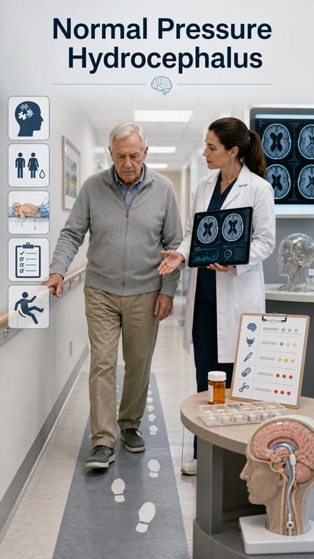
Gait impairment is often the earliest and most prominent manifestation, typically presenting as a broad-based, slow, shuffling gait with difficulty initiating movement, impaired balance, reduced stride length, and frequent falls. Patients frequently describe a sensation that their feet are “stuck to the floor,” reflecting impaired gait initiation rather than weakness.
Cognitive impairment in normal pressure hydrocephalus predominantly affects frontal and subcortical functions. Patients commonly exhibit psychomotor slowing, impaired executive function, reduced attention, diminished processing speed, and difficulties with planning and organization. Memory impairment may occur, but it is generally less severe during the early stages than in Alzheimer’s disease. Urinary symptoms usually begin with urgency and increased frequency, then progress to urge incontinence as the disease advances.
One of the greatest diagnostic challenges lies in the substantial overlap between normal pressure hydrocephalus and other disorders frequently encountered in geriatric practice. Falls, urinary urgency, executive dysfunction, white matter ischemic disease, Parkinson’s disease, atypical parkinsonian syndromes, medication-related cognitive impairment, lumbar spinal stenosis, peripheral neuropathy, depression, sleep disorders, and Alzheimer’s disease commonly coexist in older individuals. In many patients, these conditions may contribute simultaneously to functional decline, making it difficult to determine the relative contribution of ventricular enlargement to the overall clinical picture.
Neuropathological studies have found that many patients diagnosed with idiopathic normal pressure hydrocephalus also harbor concurrent Alzheimer’s disease pathology, cerebrovascular disease, or Lewy body pathology. These mixed pathologies may influence treatment response and help explain why some patients experience only partial improvement following shunt surgery. Clinicians should therefore avoid viewing normal pressure hydrocephalus as an isolated diagnosis and instead consider it within the broader context of multimorbidity and age-related neurological disease.
The clinician’s task is not merely to identify the symptom triad but also to recognize patterns that raise sufficient suspicion to warrant a comprehensive diagnostic evaluation. Initial assessment begins with a detailed clinical history, neurological examination, cognitive testing, and objective gait assessment. Particular attention should be paid to the temporal evolution of symptoms, the predominance of gait dysfunction, medication history, vascular risk factors, and evidence of alternative neurological disorders.
Neuroimaging plays a central role in establishing the diagnosis. Magnetic resonance imaging is generally preferred because it provides superior visualization of ventricular enlargement, cortical morphology, white matter changes, and alternative intracranial pathology. Imaging findings suggestive of normal pressure hydrocephalus include ventriculomegaly disproportionate to cortical atrophy, narrowing of the high-convexity sulci, enlargement of the Sylvian fissures, and disproportionately enlarged subarachnoid-space hydrocephalus (DESH). Additional imaging markers, including measurement of the Evans Index and callosal angle, may further support diagnostic confidence when interpreted alongside clinical findings.
Physiological testing also informs patient selection for surgical treatment. Large-volume cerebrospinal fluid removal via lumbar tap testing allows clinicians to assess short-term improvement in gait or cognition following drainage. External lumbar drainage over several days may provide additional prognostic information in selected patients. Objective gait measurements before and after drainage are particularly valuable. A clear objective gait response increases the likelihood of improvement after shunting, but no drainage test predicts postoperative response with certainty.
Patients demonstrating compatible clinical features, supportive imaging findings, and positive cerebrospinal fluid drainage testing should undergo evaluation by an experienced neurosurgical team for consideration of cerebrospinal fluid shunting, most commonly through ventriculoperitoneal shunt placement. Although shunt surgery offers the potential for meaningful functional improvement, appropriate patient selection remains essential to maximize benefit while minimizing surgical risks such as infection, subdural hematoma, shunt malfunction, and overdrainage.
Normal pressure hydrocephalus occupies the intersection between potentially reversible neurological dysfunction and irreversible neurodegenerative disease. Its symptoms frequently overlap with those of common disorders affecting older adults, making both underdiagnosis and overdiagnosis significant clinical concerns. Accurate diagnosis requires careful integration of clinical presentation, objective functional assessment, neuroimaging, cerebrospinal fluid testing, and multidisciplinary evaluation. By maintaining an appropriate level of clinical suspicion while applying evidence-based diagnostic criteria, clinicians can identify patients most likely to benefit from intervention.
Why NPH Is Still Missed
Idiopathic normal pressure hydrocephalus remains underrecognized despite being one of the few potentially reversible contributors to gait impairment and cognitive decline in older adults. Delayed or missed diagnosis may result in prolonged disability and lost opportunities to consider effective treatment. The difficulty lies not only in the disorder’s clinical heterogeneity but also in its overlap with more common age-related neurological and musculoskeletal conditions.
One reason iNPH is overlooked is that patients often do not present with the complete clinical triad of gait disturbance, cognitive impairment, and urinary dysfunction at the same time. These features characterize the syndrome but often develop gradually and at different stages of disease progression. Clinicians who expect all three manifestations before considering the diagnosis may therefore fail to recognize patients at earlier, potentially more treatable stages of the disease.
Gait impairment is typically the earliest and most prominent manifestation of iNPH. Patients often describe increasing difficulty with walking long before cognitive or urinary symptoms become clinically apparent. The gait is characteristically slow, broad-based, and shuffling, with reduced step height and shortened stride length. Many individuals report that their feet feel “stuck to the floor,” reflecting the magnetic gait commonly described in iNPH, although this pattern is not specific to the disorder. Balance impairment, difficulty initiating movement, impaired turning, and frequent falls commonly develop as the disease progresses. Because gait dysfunction is often the first symptom to emerge, careful assessment of mobility should play a central role in the diagnostic evaluation of older adults presenting with unexplained gait abnormalities.
Cognitive impairment in iNPH also differs from the presentation seen in many neurodegenerative dementias. Rather than exhibiting prominent episodic memory loss during the early stages, patients frequently demonstrate psychomotor slowing, reduced attention, impaired executive function, diminished planning ability, decreased mental flexibility, and apathy. Family members may describe affected individuals as moving and thinking more slowly rather than becoming overtly forgetful. These subtle cognitive changes can easily be attributed to normal aging, depression, vascular cognitive impairment, or early Alzheimer’s disease, contributing further to diagnostic delay.
Urinary dysfunction generally develops later in the disease course and often begins with relatively nonspecific lower urinary tract symptoms. Patients may initially experience urinary urgency, increased frequency, nocturia, or difficulty delaying urination. Overt urinary incontinence may become apparent as the condition progresses. Because these symptoms are highly prevalent among older adults because of benign prostatic hyperplasia, overactive bladder syndrome, pelvic floor disorders, or age-related bladder dysfunction, they are frequently investigated independently without consideration of an underlying neurological cause.
Another major factor contributing to underdiagnosis is the tendency to attribute the individual symptoms of iNPH to more common disorders affecting older adults. Gait impairment may be explained by frailty, osteoarthritis, peripheral neuropathy, cervical spondylotic myelopathy, lumbar spinal stenosis, or previous stroke. Cognitive slowing may be attributed to Alzheimer’s disease, vascular dementia, Parkinson’s disease, depression, or normal aging. Urinary symptoms are commonly attributed to urological disorders rather than neurological dysfunction.
These alternative diagnoses frequently coexist with iNPH rather than excluding it. Many older patients have multiple chronic conditions, and the presence of comorbid disease should not automatically eliminate iNPH from the differential diagnosis. Clinicians should recognize that mixed pathology is common and maintain a broad differential when evaluating patients with progressive gait dysfunction and cognitive decline.
Neuroimaging presents an additional diagnostic challenge. Ventriculomegaly, one of the hallmark imaging findings in iNPH, may be incorrectly interpreted as cerebral atrophy associated with aging or neurodegenerative disease. Conversely, patients with true cerebral atrophy may occasionally be misclassified as having hydrocephalus based solely on ventricular enlargement.
Imaging markers that increase diagnostic confidence include disproportionately enlarged ventricles relative to cortical atrophy, narrowing of the high-convexity sulci, enlargement of the Sylvian fissures, a narrowed or acute callosal angle, and the pattern known as disproportionately enlarged subarachnoid-space hydrocephalus. Nevertheless, imaging findings should always be interpreted within the broader clinical context. No single radiological feature is sufficiently sensitive or specific to independently establish or exclude the diagnosis. Likewise, the absence of a single supportive imaging marker should not preclude further evaluation when the clinical presentation strongly suggests iNPH.
Epidemiological studies suggest that iNPH may be more common than referral patterns indicate. Population-based investigations have reported widely varying prevalence estimates, largely reflecting differences in diagnostic criteria, imaging definitions, and study methodology. A prospective Swedish population study estimated the prevalence of iNPH at approximately 3.7% among individuals aged 65 years and older using modified American-European diagnostic criteria and approximately 1.5% using Japanese diagnostic criteria. These estimates should be interpreted with caution and should not be indiscriminately generalized across populations. They nevertheless reinforce that iNPH is a clinically relevant disorder that primary care physicians, geriatricians, neurologists, and neurosurgeons may encounter in routine practice.
Current Evidence: What Clinicians Should Take From It
The contemporary evidence base supports cerebrospinal fluid shunting as the primary disease-directed treatment for carefully selected patients with idiopathic normal pressure hydrocephalus. Earlier observational studies, clinical guidelines, and systematic reviews consistently reported favorable outcomes following shunt surgery, but many were limited by heterogeneous patient populations, inconsistent diagnostic criteria, variable outcome measures, and a relatively small number of randomized controlled trials. These limitations contributed to uncertainty regarding patient selection and the magnitude of treatment benefit.
More recently, higher-quality clinical evidence has strengthened confidence in shunt therapy while providing a more nuanced understanding of expected outcomes. A 2025 randomized trial provided placebo-controlled evidence on shunt effectiveness. In this randomized, double-blind study, patients who demonstrated improvement in gait velocity after cerebrospinal fluid drainage testing were selected for shunt surgery. After implantation, participants were randomly assigned to either an active low-pressure valve setting or a high-pressure placebo valve setting designed to minimize cerebrospinal fluid diversion.
At three months, participants assigned to active cerebrospinal fluid diversion had significantly greater improvement in gait velocity and Tinetti Performance Oriented Mobility Assessment scores than participants assigned to the high-pressure placebo setting. These placebo-controlled findings support a short-term benefit of active diversion for gait and balance in this selected, drainage-responsive population and reinforce the value of objective functional testing during patient selection.
In contrast, improvements in cognitive function and urinary symptoms were less definitive. Patients receiving active shunting did not demonstrate statistically significant short-term improvements in Montreal Cognitive Assessment scores or overactive bladder questionnaire outcomes compared with the placebo group. These findings suggest that although cognitive and urinary improvements may occur in some patients, these domains are generally less predictable than improvements in gait and balance. The degree of benefit may depend on disease duration, underlying neurodegenerative comorbidity, baseline severity, and the presence of irreversible neuronal injury before treatment.
The randomized trial also provided information regarding procedural safety. Patients receiving active cerebrospinal fluid diversion experienced higher rates of subdural bleeding and positional headaches, complications that are recognized risks of shunt overdrainage. Falls were more frequent among patients assigned to the placebo valve setting, although the trial did not establish the mechanism responsible for this difference. These findings emphasize that while shunt surgery offers meaningful clinical benefits, it also requires careful postoperative monitoring, individualized valve adjustment, and ongoing management to minimize complications.
Collectively, the current evidence refines the expectations clinicians should communicate to patients and their families. Cerebrospinal fluid shunting should not be presented as a universal cure for the complete clinical triad of iNPH. Rather, it should be viewed as a targeted intervention capable of producing meaningful improvements in gait, balance, mobility, and functional independence in carefully selected individuals. Improvements in cognition and urinary function remain possible but are generally more variable and less predictable than motor outcomes.
These findings also reinforce the need for careful patient selection. Diagnostic evaluation should integrate clinical presentation, neuroimaging, objective gait assessment, cerebrospinal fluid drainage testing, and multidisciplinary clinical judgment rather than relying on any single diagnostic criterion. As the evidence base continues to evolve, research should focus on refining predictive biomarkers, optimizing patient-selection algorithms, improving shunt technology, and identifying factors associated with long-term neurological recovery.
Clinicians should maintain an appropriate index of suspicion for iNPH in older adults presenting with otherwise unexplained gait impairment. Earlier recognition may improve the opportunity for treatment, but outcomes depend on multiple factors, including patient selection, symptom duration and severity, comorbid neurological disease, frailty, and response to CSF drainage.
Clinical Phenotype
The gait disorder is usually the most informative feature. Patients may have a broad-based, short-stepped, slow, magnetic, or “feet stuck to the floor” gait. Turning is often impaired. Falls, start hesitation, reduced step height, and difficulty initiating walking may be prominent.
The cognitive profile is often frontal-subcortical. Slowed processing, impaired attention, reduced mental flexibility, apathy, and executive dysfunction may predominate. A predominantly amnestic syndrome with a prominent hippocampal pattern should raise concern for Alzheimer’s disease, although mixed pathology is common in older adults.
Urinary symptoms usually include urgency, frequency, nocturia, or urge incontinence. Isolated urinary incontinence without gait impairment is rarely sufficient to support a diagnosis of NPH. Medication effects, benign prostatic hyperplasia, overactive bladder, urinary tract infection, diuretics, hyperglycemia, heart failure, sleep apnea, and pelvic floor disorders may all complicate interpretation.

Table 1. Clinical Clues That Should Trigger NPH Evaluation
| Domain | Findings That Increase Suspicion | Common Pitfalls |
|---|---|---|
| Gait | Broad-based, short-stepped, magnetic gait; impaired turning; unexplained falls | Attributing all gait decline to aging, arthritis, neuropathy, or spine disease |
| Cognition | Slowed processing, apathy, attention or executive dysfunction | Labeling as Alzheimer disease without assessing gait and imaging |
| Urinary | Urgency, frequency, or urge incontinence within a gait-predominant syndrome | Treating bladder symptoms alone with anticholinergics |
| Imaging context | Ventriculomegaly with a supportive NPH pattern | Mistaking DESH for atrophy or treating ventriculomegaly alone as diagnostic |
Diagnostic Approach
The diagnosis of iNPH should be built from the clinical syndrome, brain imaging, exclusion of major mimics, and CSF drainage or other physiologic testing when appropriate. No single component is sufficient in isolation.
Step 1: Recognize the Pattern
Clinicians should suspect NPH in an older adult with progressive gait impairment plus cognitive decline or urinary symptoms, especially when the gait deficit is disproportionate to musculoskeletal findings. The timeline is useful. Gait-first disease is more typical than cognition-first disease. Rapid cognitive decline, seizures, fever, acute encephalopathy, focal deficits, or headache with papilledema should prompt urgent evaluation for other causes.
Step 2: Review Medications and Confounders
A medication review is not peripheral. Anticholinergic drugs can worsen cognition, constipation, urinary retention, blurred vision, and fall risk. Benzodiazepines, Z-drugs, opioids, sedating antihistamines, antipsychotics, gabapentinoids, and skeletal muscle relaxants may worsen balance or cognition. Dopamine-blocking drugs such as metoclopramide, prochlorperazine, and antipsychotics can cause or worsen drug-induced parkinsonism.
For pharmacists and prescribers, the key point is not to “deprescribe away” true NPH. It is to reduce noise in the evaluation. When feasible, potentially impairing medications should be minimized before formal gait or cognitive testing, while avoiding abrupt discontinuation of drugs that require tapering.
Step 3: Obtain Brain Imaging
MRI is preferred when available because it better characterizes ventricles, sulci, white matter disease, infarcts, mass lesions, and alternative diagnoses. CT can identify ventriculomegaly and may be sufficient for initial recognition when MRI is unavailable or contraindicated.
Supportive imaging findings include ventriculomegaly disproportionate to sulcal atrophy, an Evans Index greater than 0.3, a narrowed callosal angle, enlarged Sylvian fissures, tight high-convexity sulci, and disproportionately enlarged subarachnoid-space hydrocephalus, often called DESH. These findings support the diagnosis but do not prove shunt responsiveness. The Evans Index is a marker of ventriculomegaly rather than a specific diagnostic test for iNPH. Imaging must be interpreted in the context of the clinical syndrome.
Step 4: Perform Objective Baseline Testing
Before CSF drainage testing, clinicians should obtain objective baseline measures. Useful options include gait speed, Timed Up and Go, Tinetti score, stride length, turning assessment, standardized fall history, MoCA or similar cognitive screening, functional history from an informant, and urinary symptom assessment.
Consistency in measurement is essential. The same measures should be repeated after CSF removal and after shunting when performed. Objective measurement reduces the risk of overinterpreting a subjective impression.
Step 5: Use CSF Drainage Testing Thoughtfully
Large-volume lumbar puncture, often called the CSF tap test, is widely used. Many centers remove approximately 30–50 mL of CSF, then reassess gait and cognition over hours to days. A clearly positive tap test increases the likelihood of shunt response. A negative or equivocal test does not fully exclude iNPH or the possibility of improvement after shunting, especially when clinical and imaging findings remain compelling.
External lumbar drainage may be used in specialized centers when the tap test is equivocal or when greater prognostic confidence is needed. It is more resource-intensive and carries risks that include infection, headache, overdrainage, and hospitalization-related complications. Lumbar infusion testing and intracranial pressure monitoring are used in some centers but are less available in routine North American practice.
Table 2. Practical Diagnostic Pathway for Suspected iNPH
| Step | What to Do | Why It Matters |
|---|---|---|
| 1. Screen clinically | Look for gait-predominant decline plus cognitive or urinary symptoms | The triad is often incomplete |
| 2. Reduce confounding | Review anticholinergics, sedatives, dopamine blockers, opioids, and alcohol use | Drugs can mimic or worsen gait and cognitive findings |
| 3. Image the brain | MRI preferred; CT acceptable for initial recognition | Imaging supports but does not prove the diagnosis |
| 4. Measure objectively | Gait speed, Timed Up and Go, balance scale, cognitive measures, and urinary measures | Reduces subjective interpretation |
| 5. Test CSF response | Tap test or specialized drainage testing | Helps estimate the likelihood of shunt response |
| 6. Refer selectively | Neurology, neurosurgery, neuroradiology, geriatrics, and rehabilitation | Decisions often require multidisciplinary review |
Therapeutic Considerations
There is no established medication that reverses iNPH. Cholinesterase inhibitors, memantine, bladder medications, stimulants, or medications used for gait-related symptoms should not be presented as disease-directed therapy for iNPH. They may be used for comorbid conditions when appropriate, but they do not replace CSF diversion in patients with shunt-responsive disease.
The standard disease-directed treatment is CSF diversion, most commonly ventriculoperitoneal shunting. Lumboperitoneal and ventriculoatrial shunts are used in selected circumstances. Valve selection varies by center and patient factors. Programmable valves allow noninvasive adjustment, which may be useful when balancing underdrainage against overdrainage. Anti-siphon or gravitational components may be used in selected systems, although device choice should be individualized by the treating neurosurgical team.
Endoscopic third ventriculostomy has been studied, but ventriculoperitoneal shunting remains the usual standard treatment for iNPH in most clinical practice. Evidence supporting endoscopic third ventriculostomy in iNPH remains limited, and the procedure should not be considered equivalent to CSF shunting for routine iNPH care outside specialized settings.
Safety, Contraindications, and Monitoring
Shunt surgery requires an explicit discussion of benefits and harms. Important risks include infection, hemorrhage, seizures, shunt obstruction, mechanical failure, underdrainage, overdrainage, subdural hygroma or hematoma, low-pressure headache, abdominal complications, need for revision, and complications related to anesthesia or frailty.
Active infection along the planned shunt tract or at the intended distal site generally warrants deferral of implantation until the infection has been treated. Antiplatelet and anticoagulant therapy require individualized perioperative planning. The risk of interrupting antithrombotic therapy must be weighed against procedural bleeding risk, with neurosurgery and the clinician managing the antithrombotic indication involved in the decision.
Postoperative monitoring should be structured. Patients and caregivers need clear instructions about symptoms of underdrainage, overdrainage, infection, and shunt malfunction. Worsening headache, vomiting, fever, erythema or tenderness along the shunt tract, new neurological deficits, acute confusion, gait deterioration, recurrent falls, or positional headache should prompt urgent clinical evaluation.
Programmable shunts introduce a specific safety issue. MRI and other strong magnetic exposures may alter some valve settings. Patients should know the shunt model, carry device information, and inform radiology teams before MRI. Valve settings should be verified after MRI or other magnetic exposure when recommended by the device manufacturer or treating neurosurgical team. Clinicians should not assume that all programmable valves behave identically.
Table 3. Shunt-Related Safety Issues for Clinicians
| Issue | Clinical Relevance | Practical Safeguard |
|---|---|---|
| Infection | May require antimicrobial treatment and shunt revision or removal | Monitor fever, wound changes, tract redness, or tenderness |
| Overdrainage | Can cause positional headache, subdural hygroma, or subdural hematoma | Valve assessment or adjustment, imaging, and neurosurgical follow-up |
| Underdrainage or obstruction | Hydrocephalus symptoms may persist or recur | Reassess gait, cognition, imaging, and shunt function |
| MRI or magnets | Some programmable valves may undergo an unintended setting change | Document valve type and verify settings as indicated |
| Antithrombotics | Bleeding risk must be balanced against thrombotic risk | Develop an individualized perioperative plan |
| Falls | May improve after treatment but persist because of comorbidities | Rehabilitation, home-safety interventions, and assistive devices |
Clinical Implications for Pharmacists and Prescribers
Pharmacists can strengthen the NPH workup. Medication burden often clouds the clinical picture, and many patients being evaluated for NPH are exposed to drugs that impair attention, balance, continence, or blood pressure stability.
A high-yield review includes anticholinergic burden, benzodiazepines, Z-drugs, opioids, skeletal muscle relaxants, sedating antihistamines, antipsychotics, dopamine-blocking antiemetics, gabapentinoids, antihypertensive overtreatment, hypoglycemia-inducing regimens, and drugs that worsen urinary retention. Bladder anticholinergics deserve particular caution because they may reduce urgency while worsening cognition, constipation, urinary retention, or fall risk in susceptible patients.
The goal is not reflexive discontinuation. It is a cleaner diagnostic signal and a safer perioperative course. Pharmacists should identify deprescribing opportunities, tapering requirements, renal-dose considerations, medications that increase bleeding risk, clinically relevant drug interactions, fall-risk medications, and postoperative analgesic strategies that avoid unnecessary delirium or sedation.
Patient Selection and Risk Stratification
Features that generally support shunt candidacy include a compatible clinical syndrome, supportive imaging, objective gait impairment, acceptable procedural risk, and improvement after CSF drainage. Gait-predominant patients often have the clearest short-term functional gains.
Selection becomes more difficult when severe dementia precedes gait impairment, imaging is dominated by atrophy rather than hydrocephalus, advanced neurodegenerative disease is evident, severe frailty limits rehabilitation, or comorbidities make surgery high-risk. None of these factors automatically rules out benefit, but they materially affect patient selection and counseling.
Patients and families should be told which outcome is most likely. The most realistic initial goal is usually better walking, safer transfers, or greater independence. Falls may decrease but can persist because of comorbid disease. Cognitive and urinary improvements may occur, but they are less reliable and may be limited by concurrent neurological or urological pathology.
Practical Approach for Clinicians
A practical NPH workflow can be summarized in one sentence: suspect it from the gait, support it with imaging, measure it objectively, test CSF responsiveness when appropriate, and counsel honestly about shunt benefit and risk.
In primary care, geriatrics, neurology, rehabilitation, urology, psychiatry, and pharmacy practice, the most important step is not to miss the pattern. A patient with progressive gait decline, ventriculomegaly, urinary urgency, and executive dysfunction deserves more than a dementia label. Similarly, a patient with ventriculomegaly but no compatible syndrome should not be rushed toward shunting.
Clinicians should also avoid anchoring after a single diagnosis. A patient can have iNPH plus Alzheimer’s disease, iNPH plus vascular disease, or iNPH plus spinal stenosis. The presence of one disorder does not exclude another. The central question is whether treating abnormal CSF dynamics is sufficiently likely to improve a function the patient values to justify the procedural risk.
Limitations of the Evidence
Although the evidence supporting the diagnosis and management of normal pressure hydrocephalus has expanded considerably, several limitations continue to affect the interpretation and application of current findings. Variability in study design, patient selection, diagnostic criteria, outcome measures, and follow-up duration makes direct comparison across studies difficult and limits the generalizability of many reported outcomes.
One challenge is variation among diagnostic frameworks and in how those frameworks are applied. Studies have used different combinations of clinical symptoms, neuroimaging findings, cerebrospinal fluid drainage tests, and physiological measurements to establish a diagnosis of normal-pressure hydrocephalus. Patient populations included in clinical trials therefore often differ substantially with respect to disease severity, symptom duration, comorbid neurodegenerative disorders, and the estimated likelihood of responding to surgical intervention. This heterogeneity complicates efforts to identify the patients most likely to benefit from treatment.
Outcome assessment also remains inconsistent across the literature. Studies employ different gait scales, cognitive assessments, urinary symptom questionnaires, quality-of-life instruments, and definitions of clinical improvement. Some focus primarily on changes in gait velocity, while others prioritize cognitive performance or functional independence. Response to treatment may be defined by absolute score changes, percentage improvement, or clinician judgment.
Differences in follow-up intervals further complicate interpretation, with some studies reporting outcomes only a few months after intervention and others evaluating patients over several years. The absence of standardized outcome measures limits meaningful comparisons across studies and complicates evidence synthesis.
Many earlier investigations were observational and therefore subject to selection bias, referral bias, and residual confounding. Patients referred to specialized neurosurgical centers may differ markedly from those encountered in general neurology or primary care settings, potentially leading to overestimation of treatment success when results are generalized. Retrospective analyses also frequently lack standardized assessment protocols and may incompletely account for coexisting neurological disorders that influence outcomes.
The randomized controlled trial published in 2025 represents an important methodological advance by providing placebo-controlled evidence on shunt therapy. Nevertheless, its findings should be interpreted within the context of its study design. Participants were limited to individuals who demonstrated improvement following cerebrospinal fluid drainage testing, representing a selected population with a greater estimated likelihood of benefiting from shunt surgery.
Clinical outcomes were evaluated over a relatively short three-month period, limiting conclusions regarding the long-term durability of treatment effects, delayed complications, and progressive cognitive outcomes. These factors should discourage overgeneralizing the trial’s findings to all patients with suspected normal-pressure hydrocephalus.
Neuroimaging continues to play a central role in diagnosis, yet currently available imaging markers remain imperfect predictors of treatment response. Ventriculomegaly, disproportionately enlarged subarachnoid-space hydrocephalus, narrowed high-convexity sulci, a narrowed or acute callosal angle, and periventricular white matter changes may all contribute useful diagnostic information. However, no single imaging feature, and no imaging-only combination currently validated for routine clinical use, can determine shunt responsiveness by itself.
A 2026 study of three-dimensional ventricular morphology reported only modest discrimination under repeated cross-validation, further supporting the investigational status of imaging-only prediction models. Imaging should therefore be interpreted within the broader clinical context rather than used as an isolated determinant of management decisions.
Biomarker research offers another potential avenue for improving diagnostic accuracy. Cerebrospinal fluid biomarkers associated with Alzheimer’s disease, including amyloid beta and tau proteins, may assist in identifying patients with concurrent neurodegenerative pathology that could influence prognosis. However, these biomarkers have not demonstrated sufficient predictive performance to determine shunt eligibility or accurately predict postoperative outcomes when used alone. Additional validation is required before they can be routinely incorporated into clinical decision-making.
Emerging technologies such as digital gait analysis, automated neuroimaging interpretation, wearable motion sensors, and machine-learning-based predictive models have generated considerable interest. These approaches may improve the objective quantification of functional impairment or support individualized assessment. Despite encouraging preliminary findings, they remain investigational. Their integration into routine clinical practice will require prospective validation, external replication across diverse populations, and evidence that their use improves clinically meaningful outcomes.
Future Directions
The future of normal pressure hydrocephalus management should focus on improving early recognition, standardizing diagnostic pathways, refining the prediction of shunt responsiveness, and enhancing long-term postoperative care. Although advances in imaging, biomarkers, and computational analysis continue to expand diagnostic capabilities, improvements in patient outcomes will also depend on optimized clinical workflows and multidisciplinary collaboration.
Earlier identification may improve the opportunity to consider treatment. Many individuals experience progressive gait impairment, cognitive decline, and urinary dysfunction for months or years before receiving an accurate diagnosis. Because these symptoms overlap with parkinsonian syndromes, Alzheimer’s disease, vascular dementia, and age-related functional decline, normal pressure hydrocephalus remains underrecognized in many healthcare settings. Earlier referral and evaluation may allow intervention before disability becomes less reversible, although the magnitude of this effect remains difficult to quantify.
Future clinical research should prioritize standardized outcome measurement across institutions and studies. Uniform assessment tools for gait performance, balance, cognition, urinary symptoms, functional independence, caregiver burden, and health-related quality of life would facilitate comparison between studies and strengthen the evidence base for treatment recommendations. Longer follow-up periods are equally important for evaluating the durability of shunt benefit, identifying delayed complications, and understanding the temporal patterns of cognitive and urinary improvement, which may evolve differently from gait recovery.
Advances in biomarker discovery may further improve patient selection. Ongoing research into blood-based and cerebrospinal fluid biomarkers seeks to distinguish shunt-responsive idiopathic normal pressure hydrocephalus from neurodegenerative disorders with similar clinical presentations. Future biomarker panels may improve diagnostic confidence, help predict treatment response, and identify patients with mixed pathology who require individualized counseling. However, these approaches must demonstrate clear clinical utility before widespread adoption.
Artificial intelligence and advanced imaging analysis also represent areas of investigation. Automated identification of ventriculomegaly patterns, quantitative assessment of ventricular morphology, and machine-learning algorithms that integrate imaging, clinical, and laboratory variables may improve diagnostic consistency or reduce interobserver variability. Prospective studies are needed to determine whether these technologies improve diagnostic accuracy or patient outcomes beyond conventional clinical evaluation.
Healthcare systems need not wait for future technological advances to improve detection and management. Radiologists can highlight imaging features suggestive of normal pressure hydrocephalus when ventriculomegaly and characteristic anatomical patterns are present. Primary care physicians and geriatricians can incorporate gait-focused questions and mobility assessment into evaluations of older adults presenting with falls, slowed walking, or declining mobility.
Pharmacists can assist in identifying medications that contribute to gait instability, cognitive impairment, or urinary symptoms, thereby reducing diagnostic confounding. Neurologists and neurosurgeons can collaborate to establish standardized protocols for cerebrospinal fluid tap testing, referral criteria, surgical decision-making, postoperative valve adjustment, and long-term clinical follow-up.
Normal pressure hydrocephalus should not be viewed simply as the classic example of a reversible dementia. It is a potentially treatable neurological syndrome in which gait dysfunction and balance impairment often provide the earliest and most predictable opportunity for clinical improvement. Although cognitive and urinary symptoms are important components of the disease, improved mobility often produces the most meaningful gains in independence and quality of life and may reduce caregiver burden.
Accurate diagnosis requires a systematic approach that integrates clinical assessment, recognition of the characteristic symptom pattern, exclusion of alternative neurological disorders, objective functional testing, supportive neuroimaging findings, and thoughtful use of cerebrospinal fluid drainage procedures. No single clinical feature, imaging marker, or laboratory test is sufficient in isolation. Diagnosis instead relies on synthesizing multiple sources of information while considering each patient’s overall clinical context.
Clinicians should avoid two equally problematic extremes. Normal pressure hydrocephalus should not be dismissed as an exceptionally rare condition that is unlikely to be encountered in routine practice. At the same time, shunt surgery should not be presented as a universal cure for cognitive decline or mobility impairment. Careful patient selection remains essential, as treatment response varies according to disease stage, comorbid neurological pathology, frailty, procedural risk, and individual patient characteristics.
The most effective clinical strategy is based on precision and evidence. Clinicians should identify patients with the highest likelihood of benefiting from intervention, use objective measures to document clinical change, reduce diagnostic uncertainty by addressing medication-related and comorbid confounders, provide realistic counseling about expected benefits and potential complications, and maintain structured postoperative surveillance. This balanced approach can turn recognition of a frequently overlooked, potentially reversible contributor to disability into an opportunity to preserve mobility, independence, and quality of life for appropriately selected patients.
References
Adams, R. D., Fisher, C. M., Hakim, S., Ojemann, R. G., & Sweet, W. H. (1965). Symptomatic occult hydrocephalus with “normal” cerebrospinal fluid pressure: A treatable syndrome. The New England Journal of Medicine, 273, 117–126. https://doi.org/10.1056/NEJM196507152730301. PMID: 14303656
American Geriatrics Society Beers Criteria Update Expert Panel. (2023). The American Geriatrics Society 2023 updated AGS Beers Criteria® for potentially inappropriate medication use in older adults. Journal of the American Geriatrics Society, 71(7), 2052–2081. https://doi.org/10.1111/jgs.18372. PMID: 37139824
Andersson, J., Rosell, M., Kockum, K., Lilja-Lund, O., Söderström, L., & Laurell, K. (2019). Prevalence of idiopathic normal pressure hydrocephalus: A prospective, population-based study. PLOS ONE, 14(5), e0217705. https://doi.org/10.1371/journal.pone.0217705. PMID: 31141553
Carlsen, J. F., Munch, T. N., Hansen, A. E., Hasselbalch, S. G., & Rykkje, A. M. (2022). Can preoperative brain imaging features predict shunt response in idiopathic normal pressure hydrocephalus? A PRISMA review. Neuroradiology, 64, 2119–2133. https://doi.org/10.1007/s00234-022-03021-9
Food and Drug Administration. (2018). Risks of CSF shunts. Retrieved July 13, 2026, from https://www.fda.gov/medical-devices/cerebral-spinal-fluid-csf-shunt-systems/risks-csf-shunts
Food and Drug Administration. (2019). Magnetic field interference with programmable CSF shunts. Retrieved July 13, 2026, from https://www.fda.gov/medical-devices/cerebral-spinal-fluid-csf-shunt-systems/magnetic-field-interference-programmable-csf-shunts
Food and Drug Administration. (2019). Information for clinicians about magnetic field interference. Retrieved July 13, 2026, from https://www.fda.gov/medical-devices/cerebral-spinal-fluid-csf-shunt-systems/information-clinicians-about-magnetic-field-interference
Giordan, E., Palandri, G., Lanzino, G., Murad, M. H., & Elder, B. D. (2019). Outcomes and complications of different surgical treatments for idiopathic normal pressure hydrocephalus: A systematic review and meta-analysis. Journal of Neurosurgery, 131(4), 1024–1036. https://doi.org/10.3171/2018.5.JNS1875. PMID: 30497150
Halperin, J. J., Kurlan, R., Schwalb, J. M., Cusimano, M. D., Gronseth, G., & Gloss, D. (2015). Practice guideline: Idiopathic normal pressure hydrocephalus: Response to shunting and predictors of response. Neurology, 85(23), 2063–2071. https://doi.org/10.1212/WNL.0000000000002193
Hamilton, M. G., Tullberg, M., Toma, A. K., Williams, M. A., & Klinge, P. M. (2025). Guidelines for diagnosis and management of idiopathic normal pressure hydrocephalus. Neurosurgery Clinics of North America, 36(2), 199–205. https://doi.org/10.1016/j.nec.2024.12.006. PMID: 40054973
Ishikawa, M., Hashimoto, M., Mori, E., Kuwana, N., & Kazui, H. (2012). The value of the cerebrospinal fluid tap test for predicting shunt effectiveness in idiopathic normal pressure hydrocephalus. Fluids and Barriers of the CNS, 9, Article 1. https://doi.org/10.1186/2045-8118-9-1. PMID: 22239832
Luciano, M. G., Williams, M. A., Hamilton, M. G., et al. (2025). A randomized trial of shunting for idiopathic normal-pressure hydrocephalus. The New England Journal of Medicine, 393, 2198–2209. https://doi.org/10.1056/NEJMoa2503109. PMID: 40960253
Nakajima, M., Yamada, S., Miyajima, M., et al. (2021). Guidelines for management of idiopathic normal pressure hydrocephalus, third edition: Endorsed by the Japanese Society of Normal Pressure Hydrocephalus. Neurologia Medico-Chirurgica, 61(2), 63–97. https://doi.org/10.2176/nmc.st.2020-0292. PMID: 33455998
Penkauskas, A., Kuukkanen, E., Lipponen, A., et al. (2026). Brain ventricle morphology markers in predicting shunt surgery outcome in idiopathic normal-pressure hydrocephalus. Fluids and Barriers of the CNS, 23, Article 56. https://doi.org/10.1186/s12987-026-00788-4
Relkin, N., Marmarou, A., Klinge, P., Bergsneider, M., & Black, P. M. (2005). Diagnosing idiopathic normal-pressure hydrocephalus. Neurosurgery, 57(3 Suppl), S4–S16. https://doi.org/10.1227/01.NEU.0000168185.29659.C5. PMID: 16160425
Toma, A. K., Papadopoulos, M. C., Stapleton, S., Kitchen, N. D., & Watkins, L. D. (2013). Systematic review of the outcome of shunt surgery in idiopathic normal-pressure hydrocephalus. Acta Neurochirurgica, 155(10), 1977–1980. https://doi.org/10.1007/s00701-013-1835-5. PMID: 23975646
Williams, M. A., & Relkin, N. R. (2013). Diagnosis and management of idiopathic normal-pressure hydrocephalus. Neurology: Clinical Practice, 3(5), 375–385. PMID: 24175154
Recent Articles


Integrative Perspectives on Cognition, Emotion, and Digital Behavior

Sleep-related:
Longevity/Nutrition & Diet:
Philosophical / Happiness / Social:
Other:
Modern Mind Unveiled
Developed under the direction of David McAuley, Pharm.D., this collection explores what it means to think, feel, and connect in the modern world. Drawing upon decades of clinical experience and digital innovation, Dr. McAuley and the GlobalRPh initiative translate complex scientific ideas into clear, usable insights for clinicians, educators, and students.
The series investigates essential themes–cognitive bias, emotional regulation, digital attention, and meaning-making—revealing how the modern mind adapts to information overload, uncertainty, and constant stimulation.
At its core, the project reflects GlobalRPh’s commitment to advancing evidence-based medical education and clinical decision support. Yet it also moves beyond pharmacotherapy, examining the psychological and behavioral dimensions that shape how healthcare professionals think, learn, and lead.
Through a synthesis of empirical research and philosophical reflection, Modern Mind Unveiled deepens our understanding of both the strengths and vulnerabilities of the human mind. It invites readers to see medicine not merely as a science of intervention, but as a discipline of perception, empathy, and awareness–an approach essential for thoughtful practice in the 21st century.
The Six Core Themes
I. Human Behavior and Cognitive Patterns
Examining the often-unconscious mechanisms that guide human choice-how we navigate uncertainty, balance logic with intuition, and adapt through seemingly irrational behavior.
II. Emotion, Relationships, and Social Dynamics
Investigating the structure of empathy, the psychology of belonging, and the influence of abundance and selectivity on modern social connection.
III. Technology, Media, and the Digital Mind
Analyzing how digital environments reshape cognition, attention, and identity- exploring ideas such as gamification, information overload, and cognitive “nutrition” in online spaces.
IV. Cognitive Bias, Memory, and Decision Architecture
Exploring how memory, prediction, and self-awareness interact in decision-making, and how external systems increasingly serve as extensions of thought.
V. Habits, Health, and Psychological Resilience
Understanding how habits sustain or erode well-being-considering anhedonia, creative rest, and the restoration of mental balance in demanding professional and personal contexts.
VI. Philosophy, Meaning, and the Self
Reflecting on continuity of identity, the pursuit of coherence, and the construction of meaning amid existential and informational noise.
Keywords
Cognitive Science • Behavioral Psychology • Digital Media • Emotional Regulation • Attention • Decision-Making • Empathy • Memory • Bias • Mental Health • Technology and Identity • Human Behavior • Meaning-Making • Social Connection • Modern Mind
Video Section