Neuroinflammation in Depression: Biomarker or Target?

Abstract
Major depressive disorder (MDD) remains a leading global cause of disability, yet its underlying pathophysiology is incompletely understood. Increasing evidence implicates immune and inflammatory processes in the onset, progression, and treatment response of depression. This review synthesizes current findings on the role of neuroinflammation in MDD, evaluating whether inflammatory markers function primarily as correlational biomarkers or represent viable therapeutic targets. We integrate data from clinical cohorts, neuroimaging studies, and biomarker analyses to outline the principal biological pathways by which immune dysregulation may influence mood and cognition. In addition, the potential benefits and limitations of anti-inflammatory treatment strategies are critically appraised. We conclude by highlighting key uncertainties and future directions for translational research, with particular attention to the practical implications for clinical decision-making. A concluding FAQ section provides concise, evidence-based responses to common questions raised by frontline practitioners.
Introduction
The etiology of major depressive disorder has long been framed in terms of monoaminergic neurotransmission, dysregulated hypothalamic–pituitary–adrenal (HPA) axis function, and psychosocial stressors. Over the past two decades, however, an alternative framework has emerged: the immune–inflammation hypothesis of depression. This model posits that activated immune cells and their pro-inflammatory mediators can alter neurocircuitry, neurotransmitter metabolism, and neuroplasticity in ways that contribute to depressive symptomatology.
Support for this hypothesis derives from converging lines of evidence, including elevated circulating cytokine levels in subsets of patients, cerebrospinal fluid analyses demonstrating immune activation, neuroimaging studies showing altered microglial activity, and randomized clinical trials testing anti-inflammatory interventions. Despite these advances, questions remain regarding the strength and specificity of the association between inflammation and depression, the clinical utility of measuring inflammatory biomarkers, and the effectiveness of immunomodulatory treatments across diverse patient populations.
This review seeks to provide healthcare professionals—including psychiatrists, primary care physicians, psychologists, and advanced practice nurses—with a clear and clinically relevant synthesis of the evidence. By contextualizing recent scientific developments within the realities of patient care, we aim to inform diagnostic reasoning, therapeutic decision-making, and interdisciplinary collaboration in the management of MDD.
Neuroinflammation and Depression: Current Evidence
Peripheral markers
Most studies measure proteins in the blood. The findings that repeat across labs are:
• Higher levels of C-reactive protein (CRP) in many people with MDD.
• Higher levels of cytokines such as interleukin-6 (IL-6), tumor necrosis factor-alpha (TNF-α), and interleukin-1 beta (IL-1β).
Large pooled analyses suggest that about 25–30 % of adults with MDD show CRP values >3 mg/L, a cut-off often used in heart disease risk (Osimo et al., 2020). These values drop after recovery in some—but not all—patients, hinting that inflammation might track illness activity in a subgroup.
Central markers
Cerebrospinal fluid (CSF) investigations remain relatively limited due to the invasive nature of lumbar puncture. Nevertheless, meta-analytic evidence indicates that patients with major depressive disorder (MDD) exhibit elevated concentrations of interleukin-6 (IL-6) and interleukin-8 (IL-8) compared with healthy controls (Enache et al., 2019). Advances in neuroimaging, particularly positron emission tomography (PET), now allow in vivo assessment of microglial activation—the central nervous system’s resident immune cells. Using this approach, two independent research groups reported increased microglial binding in the anterior cingulate cortex of unmedicated individuals with MDD (Setiawan et al., 2015; Holmes et al., 2018). Although sample sizes were modest, the results parallel peripheral findings: participants with higher circulating C-reactive protein (CRP) levels also demonstrated greater PET signal intensity, reinforcing the link between systemic inflammation and central neuroimmune activation.
Genetic clues
Genome-wide studies show that common gene variants tied to CRP and IL-6 pathways overlap modestly with risk for depression (Milaneschi et al., 2017). Mendelian randomisation, a tool that uses gene variants as “natural experiments,” suggests that genetically higher IL-6 increases the odds of depression, while genetically higher CRP might slightly lower the risk (Khandaker et al., 2021). The mixed pattern hints at complex, possibly feedback, loops.
Experimental and longitudinal data
Experimental studies in healthy volunteers demonstrate that acute immune activation—for instance, following administration of typhoid vaccination or endotoxin—can induce transient depressive-like symptoms, including low mood, fatigue, and social withdrawal within hours (DellaGioia & Hannestad, 2010). In clinical populations, elevated baseline C-reactive protein (CRP) has been associated with reduced responsiveness to conventional antidepressant therapies and an increased likelihood of relapse over time (Strawbridge et al., 2015). Together, these findings support the view that inflammation is not merely a downstream consequence of depression but may play a causal role in its onset, persistence, and trajectory.

Mechanisms Linking Inflammation to Mood Changes
Cytokine effects on brain circuits
Cytokines in blood can signal the brain through several routes: crossing leaky parts of the blood–brain barrier, activating nerve endings of the vagus nerve, or via cytokine transporters. Once inside the brain, cytokines alter neurotransmitter release. IL-1β and TNF-α, for instance, reduce the firing of serotonin neurons and increase glutamate release, raising the risk of excitotoxicity. In animal models, blocking IL-1β stops stress-induced loss of pleasure-seeking behavior.
Tryptophan and kynurenine pathway
Inflammation drives the enzyme indoleamine 2,3-dioxygenase (IDO), which diverts tryptophan away from making serotonin and towards kynurenine. Kynurenine can cross into the brain, where it is turned into quinolinic acid, an NMDA receptor agonist that can damage neurons. Depressed patients with high CRP show lower plasma tryptophan and higher kynurenine/tryptophan ratios (Haroon et al., 2020).
Neuroendocrine interactions
Cytokines activate the hypothalamic-pituitary-adrenal (HPA) axis, which increases cortisol levels. Over time, this may weaken the function of glucocorticoid receptors, making it harder for the body to stop inflammation. It can lead to a cycle of stress and immune activation.
Neuroplasticity and synaptic pruning
Microglia help shape synaptic connections. When over-activated, they may prune healthy synapses. Rodent studies show that chronic stress leads to microglial activation, loss of spine density on pyramidal neurons, and depressive behavior, all reversible by microglial inhibitors.
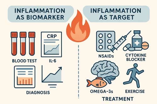
Biomarker Potential
Diagnostic aid
At present, no single inflammatory marker has enough accuracy to confirm or rule out MDD. However, a panel combining CRP, IL-6, and TNF-α reaches area-under-curve (AUC) values of 0.73–0.78 in some cohorts (Liu et al., 2021). These numbers are fair but not high enough for stand-alone tests. Importantly, raised markers also occur in obesity, smoking, infection, and autoimmune disorders, limiting specificity.
Prognostic tool
Baseline CRP over 3–5 mg/L doubles the odds of poor response to selective serotonin reuptake inhibitors (SSRIs) (Uher et al., 2014). Patients with elevated IL-6 are more likely to relapse within a year, even if they first reach remission. Thus, inflammatory panels may help stratify risk and plan closer follow-up.
Treatment matching
Early data suggest that patients with high inflammation levels may respond more effectively to specific treatments, such as bupropion, ketamine, or a combination therapy that includes an anti-inflammatory. A 2019 trial found that infliximab failed in the full sample of resistant depression, but in the subgroup with CRP >5 mg/L, the drug cut symptoms by ~50 % (Raison et al., 2013). Similar “enrichment” designs are underway with tocilizumab and minocycline.

Inflammation as a Treatment Target
Non-steroidal anti-inflammatory drugs (NSAIDs)
Adding celecoxib (400 mg/day) to an SSRI showed modest but significant extra benefit in several 6-to-8-week trials (Nielsen et al., 2019). Safety remains a concern in long-term use, given gastric and cardiac risks.
Cytokine antagonists
Monoclonal antibodies against TNF-α (infliximab) and IL-6 receptor (tocilizumab) are approved for rheumatoid arthritis. Open-label studies in MDD with high CRP suggest symptom drops within 4–6 weeks, though randomised data are sparse and costly.
Minocycline
This antibiotic crosses the blood–brain barrier and dampens microglial activation. Pooled data from five small trials show a moderate effect size (SMD ≈ –0.4) in resistant depression (Rosenblat & McIntyre, 2018). Upset stomach and skin changes are the common side effects, but the risk of resistance prompts caution.
Omega-3 fatty acids
Eicosapentaenoic acid (EPA) at doses ≥2 g/day lowers IL-6 and may have minor antidepressant effects, especially in women and those with high baseline inflammation.
Lifestyle measures
Exercise, sleep regulation, and anti-inflammatory diets (more fruit, vegetables, whole grains; less processed food) all reduce CRP by 10–20 % and improve mood scores. Though less dramatic than drugs, such steps carry a lower risk and can be sustained.
Applications and Use Cases
In current practice, guiding treatment by inflammation status is still experimental, yet some realistic scenarios are:
- A patient with resistant depression and obesity has CRP 8 mg/L; adding celecoxib for 8 weeks might be reasonable while awaiting weight-loss measures.
- An outpatient with multiple past antidepressant failures is screened; if IL-6 is high, referral to an anti-cytokine trial could be suggested.
- In relapse prevention clinics, measuring CRP every 6–12 months may flag those needing closer follow-up or early psychotherapy boosters.
Comparison with Other Models of Depression
Traditional monoamine theories hold that low serotonin or norepinephrine drives depression. Inflammation affects these transmitters upstream. Neuroplasticity models focus on brain-derived neurotrophic factor (BDNF) and synapse health; cytokines lower BDNF, linking the two views. The HPA axis model and the inflammation model are intertwined, as cytokines stimulate cortisol release and cortisol feeds back on immune cells. Rather than competing frameworks, these theories describe different layers of the same system.

Conclusion 
Challenges, Limitations, and Biases
Sampling bias is common: most studies involve small, clinic-based samples that may not represent primary care populations. Confounders like smoking, diet, and sleep are not consistently controlled. Laboratory methods differ; CRP is stable, but cytokines can stick to tube walls and degrade, causing wide lab-to-lab variation. PET ligands for microglia bind not only to activated cells but also to endothelial cells, limiting specificity. Finally, most drug trials last under three months, so the long-term safety of anti-inflammatory drugs in MDD remains unknown.
Future Directions and Recommendations
Future research should prioritize large, pragmatic clinical trials that stratify patients according to baseline inflammatory status in order to clarify treatment implications. Integrated multi-omic approaches—encompassing cytokine profiling, metabolomics, and gene expression analyses—hold promise for enhancing diagnostic precision and identifying biologically distinct subtypes of depression. Development of more accessible PET or MRI biomarkers of microglial activation would further advance translational applications. From a clinical perspective, when elevated C-reactive protein (CRP) is identified in a patient with depression, careful evaluation for underlying infection, autoimmune conditions, or metabolic syndrome is warranted, as targeted management of these comorbidities may contribute to improvements in mood and overall functioning.

Frequently Asked Questions:
- Why do some individuals with depression present with normal CRP levels? Inflammation represents only one of several etiological pathways to depressive syndromes. Genetic vulnerability, dysregulation of the hypothalamic–pituitary–adrenal (HPA) axis, and psychosocial stressors can all precipitate mood disturbance in the absence of elevated systemic inflammatory markers.
- Should I order CRP on every new depression case? There is no guideline mandating this. Yet a baseline CRP can help rule out hidden infection or autoimmunity and may guide future treatment choices.
- Are over-the-counter nonsteroidal anti-inflammatory drugs (NSAIDs) safe for self-treatment of depression? Routine use is not recommended. NSAIDs carry well-documented risks, including gastrointestinal ulceration, renal impairment, and increased cardiovascular events. Any consideration of anti-inflammatory therapy for depressive symptoms should occur within a structured treatment plan under medical supervision.
- Does elevated inflammation imply that depression is a “physical” rather than a “mental” disorder? The distinction is artificial. Immune activation can alter neural signaling and brain function, illustrating the deep interconnection between mind and body. Depression with an inflammatory component is therefore simultaneously biological and psychological. Importantly, labeling a condition as “mental” does not diminish its reality or clinical significance.
- Will anti-cytokine drugs replace antidepressants?Unlikely. They may help a subset of patients, likely in combination with standard therapies. Cost, infection risk, and screening needs limit broad use.
- Can lifestyle changes lower both inflammation and depression? Yes. Regular aerobic exercise, a balanced diet, good sleep, and avoiding tobacco lower CRP and improve mood in many studies.
References:
DellaGioia, N., & Hannestad, J. (2010). A critical review of human endotoxin administration as an experimental paradigm of depression. Neuroscience & Biobehavioral Reviews, 34(1), 130-143. https://doi.org/10.1016/j.neubiorev.2009.07.014
Enache, D., Pariante, C., & Mondelli, V. (2019). Markers of central inflammation in major depressive disorder: A systematic review and meta-analysis of studies examining cerebrospinal fluid, positron emission tomography and post-mortem brain tissue. Brain, Behavior, and Immunity, 81, 24-40. https://doi.org/10.1016/j.bbi.2019.08.027
Haroon, E., Miller, A. H., & Sanacora, G. (2020). Inflammation, glutamate, and glia: A trio of trouble in mood disorders. Neuropsychopharmacology, 45(1), 190-206. https://doi.org/10.1038/s41386-019-0441-1
Holmes, S. E., et al. (2018). Brain TSPO imaging and gray matter volume in older depressed patients treated with escitalopram. Neuropsychopharmacology, 43(5), 970-976. https://doi.org/10.1038/npp.2017.240
Khandaker, G. M., et al. (2021). Association between genetically determined interleukin-6 signalling and risk of depression: A Mendelian randomisation study. The Lancet Psychiatry, 8(8), 699-707. https://doi.org/10.1016/S2215-0366(21)00174-8
Liu, J. J., et al. (2021). Peripheral cytokine levels and response to antidepressant treatment in depression: A systematic review and meta-analysis. Psychological Medicine, 51(4), 1839-1855. https://doi.org/10.1017/S0033291719001492
Milaneschi, Y., et al. (2017). Genetic association of major depression with atypical features and obesity-related immunometabolic dysregulations. JAMA Psychiatry, 74(12), 1214-1225. https://doi.org/10.1001/jamapsychiatry.2017.3016
Nielsen, R. E., Lydholm, C. N., Hansen, H. V., & Røn-Thomsen, M. (2019). Celecoxib as augmentation therapy for patients with major depression: A meta-analysis. Journal of Affective Disorders, 246, 282-291. https://doi.org/10.1016/j.jad.2018.12.006
Osimo, E. F., et al. (2020). Inflammatory markers in depression: A systematic review and meta-analysis of 82 studies. Molecular Psychiatry, 25(2), 337-350. https://doi.org/10.1038/s41380-018-0095-y
Raison, C. L., et al. (2013). A randomized controlled trial of the tumor necrosis factor antagonist infliximab for treatment-resistant depression: The role of baseline inflammatory biomarkers. JAMA Psychiatry, 70(1), 31-41. https://doi.org/10.1001/2013.jamapsychiatry.4
Rosenblat, J. D., & McIntyre, R. S. (2018). Efficacy and tolerability of minocycline for depression: A systematic review and meta-analysis of clinical trials. Journal of Affective Disorders, 227, 219-225. https://doi.org/10.1016/j.jad.2017.11.066
Setiawan, E., et al. (2015). Association of translocator protein total distribution volume with duration of untreated major depressive disorder: A cross-sectional study. The Lancet Psychiatry, 2(9), 790-796. https://doi.org/10.1016/S2215-0366(15)00227-6
Strawbridge, R., et al. (2015). Inflammation and clinical response to treatment in depression: A meta-analysis. European Neuropsychopharmacology, 25(10), 1532-1543. https://doi.org/10.1016/j.euroneuro.2015.06.007
Uher, R., et al. (2014). An inflammatory biomarker as a differential predictor of the outcome of depression treatment with escitalopram and nortriptyline. American Journal of Psychiatry, 171(12), 1278-1286. https://doi.org/10.1176/appi.ajp.2014.14010055


