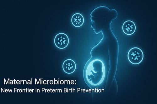
Microbiome During Pregnancy New Links to Preterm Birth Prevention

Introduction
Preeclampsia remains a major global health challenge, affecting an estimated 2 to 8 percent of pregnancies worldwide. It contributes to approximately 76,000 maternal deaths and 500,000 fetal deaths each year. This condition, characterized by new-onset hypertension and multi-organ involvement during pregnancy, significantly increases the risk of maternal morbidity and mortality. It is also a leading cause of preterm birth, which itself is responsible for nearly 1 million deaths annually and remains the foremost cause of mortality among children under the age of five worldwide. Infection is implicated in at least one-third of preterm births, underscoring the multifactorial and complex nature of these adverse outcomes.
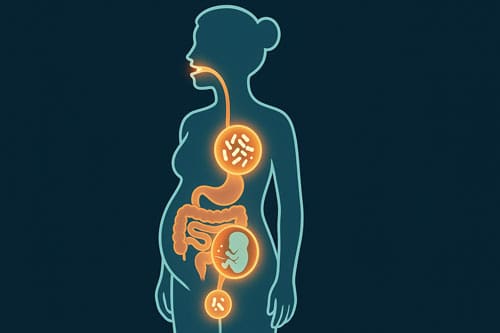
Recent research has highlighted the maternal microbiome as a critical factor influencing pregnancy outcomes. During gestation, the microbiome undergoes profound compositional shifts, particularly in the gut, vagina, and oral cavity. These microbial changes are thought to contribute not only to normal adaptations of pregnancy but also to pathological processes when dysbiosis occurs. Disturbances in microbial communities have been linked to serious complications, including preeclampsia, which can result in maternal organ damage, intrauterine growth restriction, and premature delivery. Currently, preeclampsia affects approximately 5 percent of all pregnancies and accounts for 15 percent of maternal deaths and 25 percent of neonatal deaths worldwide.
Beyond the clinical burden, the economic consequences are substantial. In the United States alone, healthcare expenditures associated with preeclamptic pregnancies are estimated at 2.18 billion dollars in the first year after delivery, encompassing both maternal and neonatal care. These costs highlight the urgent need for improved strategies to identify, prevent, and manage preeclampsia and other pregnancy complications.
An emerging body of evidence suggests that maternal gut microbiome remodeling and associated metabolic changes during pregnancy may play a pivotal role in determining maternal-fetal outcomes. Alterations in microbial metabolites, inflammatory pathways, and immune regulation could contribute to disease development and progression. At the same time, the establishment of the neonatal gut microbiota is increasingly recognized as a cornerstone of infant health, with early-life disturbances associated with heightened risks of metabolic, immunological, and developmental disorders that extend into later life.
This article explores the complex interplay between maternal microbiomes across different body sites and pregnancy outcomes, with a focus on their mechanistic links to preeclampsia and preterm birth. It also considers the potential for microbiome-targeted interventions, including dietary strategies, probiotics, prebiotics, and novel therapeutic approaches, as avenues to reduce the burden of maternal and neonatal complications. By advancing our understanding of these intricate relationships, the maternal microbiome may represent a promising frontier in improving maternal-fetal health and reducing the global impact of pregnancy-related morbidity and mortality.
Keywords: maternal microbiome, preeclampsia, preterm birth, neonatal health, pregnancy complications, microbial dysbiosis, maternal-fetal outcomes
Microbiome Changes During Pregnancy Across Body Sites
Pregnancy induces substantial alterations in the microbiome across multiple body sites, with these changes potentially influencing maternal-fetal health outcomes. These adaptations appear to follow distinct patterns throughout gestation, creating unique microbial profiles that support the physiological demands of pregnancy.
Gut microbiome during pregnancy: diversity and composition
The maternal gut microbiota undergoes remarkable transformations throughout pregnancy. While the gut microbiome composition in early pregnancy resembles that of non-pregnant women, progressive changes become evident as gestation advances [1]. Four main bacterial phyla dominate the gut microbiome: Actinobacteria, Firmicutes, Bacteroidetes, and Proteobacteria [1]. However, pregnant women exhibit higher proportions of Bifidobacterium, Proteobacteria, and Actinobacteria compared to non-pregnant controls [1].
By the third trimester, researchers observe reduced alpha diversity with simultaneously elevated beta diversity and opportunistic pathogen abundance [1]. The Firmicutes:Bacteroidetes ratio increases throughout pregnancy, mimicking patterns observed in obesity [2]. Moreover, studies demonstrate that women who gain above median gestational weight show increased bacterial diversity compared to those with lower weight gain [3]. Specifically, these women display increased abundance of Bacteroidetes (p=0.02) and other phyla (p=0.04) from baseline to follow-up [3].
Interestingly, when third-trimester gut microbiota were transplanted into germ-free mice, the animals exhibited weight gain and heightened inflammatory responses compared to those receiving first-trimester microbiota [4]. These findings suggest potential metabolic consequences of pregnancy-related microbiome shifts.
Vaginal microbiome shifts in early vs late gestation
The vaginal microbiome undergoes distinct changes across gestation periods, generally characterized by increasing Lactobacillus dominance. During pregnancy, estrogen promotes glycogen deposition in vaginal epithelium, creating favorable conditions for Lactobacillus proliferation [5]. This environment maintains a protective pH of 3.8-4.5 [1].
Research reveals that pregnant women have higher relative abundances of L. vaginalis, L. crispatus, L. gasseri, and L. jensenii compared to non-pregnant women [6]. Concurrently, pregnant women show lower abundances of 22 other non-Lactobacillus phylotypes [6]. Community state types (CSTs) based on Lactobacillus dominance show notable shifts across pregnancy. While Lactobacillus-dominated CSTs I, III, and V become more abundant as gestation progresses, diverse CSTs IV-A and IV-B decline steadily toward term [6].
Notably, the vaginal microbiota of pregnant women displays greater temporal stability than that of non-pregnant women [6]. However, dramatic shifts occur postpartum, with approximately 80% of women in Lactobacillus-dominant CSTs transitioning to non-Lactobacillus-dominant CSTs within weeks after delivery [5]. This postpartum period features increased diversity and growth of anaerobic species like Peptoniphilus, Prevotella, and Anaerococcus [4].
Oral and placental microbiome: emerging evidence
The healthy human oral cavity contains approximately 50-100 million bacteria from about 700 species [1]. During pregnancy, the oral microbiome undergoes compositional changes, particularly in the first trimester when bacterial numbers increase [1]. Pregnant women show greater abundance of Porphyromonas, Treponema, and Neisseria in saliva, while non-pregnant women exhibit more Veillonella and Streptococcus [1].
Hormonal changes in pregnancy promote bacterial plaque formation, potentially resulting in gingivitis during the second and third trimesters [1]. This condition has been associated with pregnancy complications including preeclampsia, preterm birth, low birth weight, and miscarriage [1].
The placental microbiome remains somewhat controversial but increasingly recognized. Studies suggest the placenta harbors a low-biomass microbiome established in early pregnancy [6]. The placental microbial communities exhibit limited diversity compared to other body sites [2]. Interestingly, the placental microbiome shows greater similarity to the oral microbiome than to the gut microbiome, particularly at higher taxonomic levels [2].
Analysis reveals that all phylotypes in the core placental microbiome are also present in maternal oral and gut microbiomes [2]. The shared core families between oral and placental microbiomes include Actinomycetaceae, Micrococcaceae, Oxalobacteraceae, Neisseriaceae, Pasteurellaceae, and Pseudomonadaceae [2]. Among shared genera are Actinomyces, Rothia, Haemophilus, and Pseudomonas [2].
The mechanism for potential oral-to-placental bacterial transmission appears to involve hematogenous spread, particularly noted with periodontal disease and adverse pregnancy outcomes [2]. This connection highlights the potential systemic implications of oral microbiome alterations during pregnancy.
Host Remodeling of the Gut Microbiome and Metabolic Changes During Pregnancy
The bidirectional relationship between maternal physiology and the gut microbiome represents a fascinating aspect of pregnancy. As the mother’s body adapts to support fetal growth, these physiological changes reshape microbial communities which, in turn, influence maternal metabolic and immunological processes.
Immune tolerance and inflammation balance
Throughout pregnancy, inflammatory states fluctuate according to gestational stage—from heightened inflammation during implantation and labor to reduced levels in mid-pregnancy [7]. Although anti-inflammatory properties exist at the placental bed to protect the fetus from rejection, mucosal surfaces throughout the gastrointestinal tract simultaneously experience a low-grade inflammatory state with rising levels of pro-inflammatory cytokines and white cells as pregnancy advances [7].
This delicate inflammatory balance enables proper cooperation between the trophoblast and immune system, allowing temporal invasion of T lymphocytes, macrophages, and natural killer lymphocytes that support correct angiogenesis, nutrient transport, and protection against microorganisms [8]. Nevertheless, if this balanced inflammatory status becomes disrupted by maternal conditions such as obesity or impaired intestinal barrier function, vascular dysfunction can develop in placental tissue, potentially leading to complications like fetal growth restriction and preeclampsia [7].
The gut microbiome plays an essential role in maintaining this immune balance. Studies show that the transfer of third-trimester microbiota to germ-free mice induced significantly higher levels of the pro-inflammatory cytokines IFNγ, IL2, IL6, and TNFα compared to first-trimester microbiota [6]. Furthermore, gut microbial metabolites like short-chain fatty acids (SCFAs) influence immune regulation by binding to G-protein coupled receptors (GPRs) on immune cells and inhibiting histone deacetylase activity, thereby promoting regulatory T cells (Tregs) and suppressing inflammation [5].
Hormonal regulation of microbial communities
Reproductive hormones—especially estrogen and progesterone—orchestrate substantial physiological changes during pregnancy that directly influence gut microbial composition [9]. In fact, progesterone helps balance Th1/Th2 immune responses and regulates pro-inflammatory cytokines, creating a favorable environment for certain bacterial communities [5].
Multiple studies have confirmed the regulatory relationship between hormones and the gut microbiome. Koren et al. observed profound alterations in the gut microbiome during the third trimester when estrogens reach their maximal peak, regardless of health status [10]. Beyond direct effects on bacterial growth, estrogen and progesterone enhance the expression of tight junction components in the intestinal epithelium, thereby decreasing gut barrier permeability, bacterial translocation, and inflammation [11]. These hormones likewise influence gut transit time and contractility, possibly representing an adaptive response to enhance energy extraction from food to support pregnancy demands [11].
Intriguingly, this relationship works both ways—the gut microbiome produces β-glucuronidase, which blocks the binding of estrogen to glucuronic acid, reducing estrogen inactivation and increasing circulating levels [10]. Additionally, enterohepatic circulation plays a role in this process, as hepatically conjugated estrogens excreted in bile can be deconjugated by bacterial species with β-glucuronidase activity in the gut [10].
Impact on nutrient absorption and metabolism
The normal endocrine and metabolic functions of pregnancy are designed to secure an adequate energy supply for both the mother and the developing fetus. Within this context, pregnancy-induced changes in the maternal gut microbiota play an important role in facilitating metabolic adaptation. These microbial shifts not only influence nutrient processing but also modulate host signaling pathways that support gestational physiology.
The maternal gut microbiome affects host metabolism through several mechanisms:
- Production of short-chain fatty acids (SCFAs). Metabolites such as butyrate, acetate, and propionate regulate host metabolism, influence immune system function, and control cell proliferation. These SCFAs serve as key signaling molecules linking diet, microbiota activity, and host physiology.
- Alterations in bacterial composition and energy extraction. Pregnancy is associated with shifts in gut microbial populations that influence the efficiency of energy harvest. Firmicutes are more effective than Bacteroidetes at extracting energy from the diet, which contributes to the increased energy availability required during gestation.
- Regulation of butyrate levels. Butyrate is a critical SCFA with multiple beneficial effects, including prevention of obesity induced by high-fat diets, protection of pancreatic beta-cell function, and reduction of metabolic disturbances that may arise during pregnancy. Importantly, butyrate levels show an inverse relationship with maternal blood pressure and plasminogen activator inhibitor-1 concentrations. Clinical observations demonstrate that the abundance of butyrate-producing bacteria is significantly reduced in overweight and obese women by the sixteenth week of gestation. Furthermore, experimental evidence suggests that sodium butyrate can lower blood pressure by suppressing angiotensin II activity and inhibiting the renin-angiotensin system.
Taken together, these changes highlight the dual role of the gut microbiome as both a mediator of host energy metabolism and a regulator of maternal cardiovascular and metabolic health. Interestingly, the gut microbiota during pregnancy often displays features that resemble metabolic syndrome, such as reduced microbial diversity and increased representation of Proteobacteria. In a non-pregnant state, such alterations would be considered detrimental and associated with metabolic dysfunction. However, within the unique physiological context of pregnancy, these same changes are adaptive. They enhance nutrient availability for the fetus, support maternal energy storage, and prepare the maternal body for the substantial energetic demands of lactation.
This complex interplay underscores the remarkable adaptability of the maternal gut microbiota. By reprogramming metabolic pathways in ways that would otherwise be pathological, the microbiome ensures that both maternal and fetal nutritional needs are met during pregnancy.
Vaginal Microbiome and Its Role in Preterm Birth Risk
The vaginal microbiome emerges as a critical determinant of pregnancy outcomes, with its composition strongly linked to preterm birth (PTB) risk. Unlike gut microbiota where diversity benefits health, the vaginal environment typically functions optimally with lower diversity and Lactobacillus dominance.
Lactobacillus dominance and protective effects
Lactobacillus species, particularly L. crispatus, play a pivotal role in maintaining vaginal health during pregnancy. Women who deliver at term are more likely to exhibit L. crispatus predominance in their vaginal microbiome [12]. This species creates a protective environment through multiple mechanisms: preventing pathogen adherence via coaggregation and biofilm formation; producing antibacterial compounds like lactic acid, hydrogen peroxide, and bacteriocins; and stimulating local immune responses [13].
Among the main vaginal lactobacilli (L. crispatus, L. jensenii, L. gasseri, and L. iners), L. crispatus demonstrates the strongest protective effect against preterm birth. Research reveals that women with L. crispatus dominance have approximately 20% reduced odds of spontaneous PTB [3]. In contrast, L. gasseri shows minimal antagonistic effect against group B streptococcus colonization, while L. jensenii and L. iners demonstrate only marginal protection [13].
Throughout normal pregnancy, the vaginal microbiota remains remarkably stable with high levels of lactobacilli, creating an acidic environment (pH 3.8-4.5) that inhibits pathogen growth. This stability represents a natural defense mechanism against ascending infections that might trigger premature labor.
High-diversity CST IV and inflammation
Vaginal microbiome compositions with high species diversity, designated as community state type IV (CST IV), correlate strongly with increased PTB risk. This diverse community state often features depleted Lactobacillus populations and elevated levels of anaerobic bacteria including Gardnerella, Atopobium, Prevotella, Sneathia, and Megasphaera [14].
Studies show that alpha diversity measures (ACE, Chao1, Simpson, and Shannon indices) differ substantially between term and preterm birth groups [15]. Furthermore, integrated differential abundance analyzes reveal that 18 out of 25 genera demonstrate positive associations with PTB risk, with L. iners, Prevotella, and Gardnerella showing particularly consistent connections [14].
The inflammatory response plays a crucial role in this relationship. High-diversity vaginal communities induce elevated levels of pro-inflammatory cytokines including IL-1, IL-8, IL-16, TNF-α, and IFN-γ [16]. This chronic inflammation can lead to cervical remodeling, membrane weakening, and ultimately preterm labor. Moreover, bacterial species like Gardnerella vaginalis generate proteolytic enzymes such as sialidase and proline aminopeptidase that break down protective vaginal mucins and fetal membranes, reducing their elasticity and potentially leading to premature rupture [17].
Ethnic variations in vaginal microbiota and outcomes
The composition of the vaginal microbiome exhibits considerable variation across ethnic groups, with important implications for PTB risk assessment. Black and Hispanic women more frequently display diverse CST IV profiles with lower Lactobacillus abundance compared to Asian and Caucasian women [18]. These women also tend to have higher vaginal pH (4.7-5.0) compared to Asian or White women (pH 4.2-4.4) [17].
Interestingly, while L. crispatus dominance correlates strongly with reduced PTB risk in Caucasian populations, this association appears weaker in women of African descent [1]. Yet, recent evidence suggests that regardless of ethnicity, reduced L. crispatus abundance increases PTB risk, indicating that the disparity in PTB rates may partly stem from the lower prevalence of L. crispatus-dominated microbiomes among Black women [3].
Beyond genetics, environmental and social factors also influence these ethnic variations. Practices like vaginal douching, more common among Black women, can disrupt healthy microbiome balance [3]. Additionally, structural racism may impact vaginal health through dietary access, environmental exposures, and chronic stress, which doubles the risk of bacterial vaginosis [18].
These findings underscore the need for personalized approaches to PTB prevention that consider both microbial composition and sociodemographic factors influencing vaginal health during pregnancy.
Gut Microbiome and Systemic Inflammation in Pregnancy
Beyond localized microbiome changes, systemic effects of gut microbial shifts throughout pregnancy warrant close examination. Research increasingly connects maternal gut flora alterations with inflammatory processes that may influence pregnancy outcomes, even in distant tissues and organs.
Proteobacteria and Actinobacteria shifts
Longitudinal studies reveal that maternal gut microbiome composition changes markedly between trimesters, with beta-diversity expanding dramatically from first to third trimester [4]. Most notably, relative abundances of both Proteobacteria and Actinobacteria increase substantially as pregnancy progresses [4]. Researchers observed that pregnant women undergo a decrease in alpha diversity coupled with this taxonomic shift [4].
The growth in Proteobacteria and Actinobacteria appears to serve both protective and potentially problematic functions through proinflammatory mechanisms [19]. Initially, these bacterial shifts facilitate metabolic adaptations necessary for pregnancy. Nevertheless, excessive increases might trigger pathological responses. Koren et al. demonstrated this phenomenon by transplanting third-trimester microbiota into germ-free mice, which subsequently increased fat deposition, inflammation, and insulin insensitivity compared to first-trimester microbiota [4].
Intriguingly, maternal race correlates with bacterial composition, as one study found Proteobacteria was associated with earlier gestation (≤ 21 weeks) and Black race [4]. Since both early gestation and Black race correspond with heightened pro-inflammatory states, this bacterial shift may represent an underlying mechanism connecting microbial changes with inflammation.
LPS and TMAO as pro-inflammatory metabolites
Bacterial metabolites represent critical mediators between gut microbiome changes and systemic inflammation. Chief among these is lipopolysaccharide (LPS), an endotoxin present in the cell walls of gram-negative bacteria. Increased epithelial permeability facilitates LPS penetration into circulation, resulting in what researchers term “metabolic endotoxemia” [19].
Once in circulation, LPS stimulates the immune system via toll-like 4 receptors on intestinal epithelium, thereby activating numerous cell-signaling pathways that induce inflammatory responses and cytokine secretion [20]. These pathways include activation of:
- Myeloid differentiation factor 88 (MyD-88)
- IL-1 receptor-associated kinase (IRAK)
- TNF receptor-associated factor 6 (TRAF6)
- TGF-β-activated kinase1 (TAK1) [21]
Another key metabolite, trimethylamine N-oxide (TMAO), emerges as a potential risk factor for preeclampsia. Certain gut bacteria produce trimethylamine lyase, which converts dietary components like choline and L-carnitine to trimethylamine (TMA), subsequently oxidized to TMAO by flavin monooxygenases in the liver [5]. Presently, research indicates that both fecal and plasma TMAO concentrations are higher in patients with preeclampsia than in healthy controls [5].
TMAO promotes oxidative stress and inflammation through NF-kB pathway activation, increasing expression of inflammatory cytokines including IL-1β, IL-6, and TNF-α [22]. Furthermore, TMAO directly inhibits nitric oxide production in endothelial cells, leading to vasoconstriction and placental ischemia [22].
Gut barrier integrity and immune activation
Intestinal permeability plays a pivotal role in linking gut microbiota with systemic inflammation. Throughout pregnancy, several physiological changes affect barrier function. As the uterus expands, it compresses abdominal organs, affecting intestinal motility. Simultaneously, elevated progesterone levels relax smooth muscles, slowing intestinal movement and delaying gastric emptying [23].
When barrier integrity becomes compromised, bacterial components like LPS enter circulation, triggering toll-like receptor activation and subsequent inflammatory cascades. This process creates a feedback loop wherein inflammation further increases barrier permeability, allowing additional bacterial translocation [24].
Animal studies confirm these mechanisms, as mice receiving fecal transplants from preeclamptic women displayed noticeably higher oxidative stress damage in placentas, marked by increased malondialdehyde content and decreased activity of antioxidant enzymes such as superoxide dismutase and catalase [5]. In addition, these animals exhibited higher serum levels of inflammatory factors, including interleukin-6, tumor necrosis factor-α, and interleukin-1β [5].
Maternal Microbiome and Offspring Allergy and Asthma Risk
Emerging research demonstrates the profound impact of maternal microbiome composition on offspring health outcomes beyond pregnancy complications. The intricate relationship between maternal microbial communities and infant immune development represents a critical window for allergy and asthma prevention.
Vertical transmission of maternal gut microbiota
Multiple pathways facilitate maternal-to-infant bacterial transmission. Recent studies confirm that maternal intestinal, vaginal, and oral microbes contribute to fetal gut colonization, with maternal gut microbiota representing the most influential source [25]. Vertical transmission occurs through three primary routes: prenatal transplacental transfer, birth canal exposure, and postnatal transmission via breastfeeding.
Mode of delivery critically affects this transmission. Vaginal delivery enables direct exposure to maternal vaginal and gut bacteria, whereas cesarean section disrupts this natural process [25]. Interestingly, after administration of Lactobacillus rhamnosus to pregnant women in the third trimester, the bacterium was detected in infant stool samples regardless of delivery mode [9]. This finding challenges traditional understanding of bacterial colonization pathways.
Bacterial strains with specific genetic and metabolic capabilities demonstrate enhanced transmissibility. Bifidobacterium strains containing genes for human milk oligosaccharide degradation exhibit higher transfer rates and persistence in infant guts [25]. These strains comprise approximately 11% of early colonizers and persist throughout the first year of life [25].
Early-life immune programming via SCFAs
Short-chain fatty acids (SCFAs)—primarily acetate, propionate, and butyrate—serve as crucial metabolites mediating microbiome-immune interactions. These bacterial fermentation products exert potent immunomodulatory effects through multiple mechanisms.
SCFAs influence immune development by inhibiting histone deacetylase (HDAC), particularly in regulatory T cells (Tregs). By inhibiting HDAC, butyrate keeps DNA transcriptionally active, upregulating genes involved in mucus production and tight junction formation [10]. This process strengthens intestinal barrier function, reducing permeability that otherwise promotes allergic sensitization.
Animal studies provide compelling evidence for SCFA’s protective effects. Pregnant mice fed high-fiber diets experienced gut microbiome alterations with increased SCFA production, leading to suppression of allergic airway disease in their offspring [26]. This protection stems from elevated SCFA levels promoting Treg development [26]. Correspondingly, maternal acetate supplementation attenuated offspring allergic disease through epigenetic modification at the FOXP3 promoter [27].
Associations with asthma and allergic diseases
Epidemiological evidence links maternal microbiome composition to offspring allergy risk. Farm animal exposure during pregnancy significantly reduces childhood allergy incidence, with immunological tolerance already present in cord blood [11]. Mechanistically, this exposure impacts neonatal Treg quantity and function, contributing to reduced asthma rates [11].
Maternal antibiotic use during pregnancy demonstrates opposite effects. Danish birth cohort data showed prenatal antibiotic exposure increased atopic dermatitis odds at 18 months in infants of atopic mothers [11]. Similarly, maternal antibiotic use correlated with 1.3-fold higher asthma risk in children aged 2-10 years [11]. Notably, third-trimester antibiotic exposure appears particularly influential [11].
Specific bacterial taxa exhibit protective associations. Lower fecal abundance of Prevotella copri in mothers correlates with increased food allergy risk in their infants [27]. Furthermore, maternal vaginal clusters dominated by L. jensenii demonstrate immunosuppressive effects on fetal antigen-presenting cells [28], potentially promoting tolerance against allergens.
Human milk provides another transmission pathway for protective bacteria and their metabolites. Research indicates infants with atopic dermatitis received markedly lower levels of SCFAs, particularly acetate, in human milk compared to healthy controls [10]. Even modest concentrations of milk-derived SCFAs can shift immune responses from pro-inflammatory to anti-inflammatory states [10].

Mechanistic Links Between Microbiome and Preterm Birth
Recent molecular investigations reveal multiple pathways through which microbial dysbiosis triggers preterm labor and delivery. These mechanisms extend beyond simple microbial presence, involving complex interactions between microbes, their metabolites, and maternal-fetal immune responses.
Microbial metabolites and cytokine signaling
The cervicovaginal metabolome reflects dynamic host-microbiota interactions that can trigger inflammatory cascades leading to premature birth. Women who deliver preterm show distinctive metabolite profiles associated with specific bacterial communities. In particular, bacterial vaginosis-associated bacteria produce metabolites like 2-hydroxyisovalerate and γ-hydroxybutyrate that correlate with clinical symptoms [29]. Furthermore, elevated 12-hydroxyeicosatetraenoic acid (12-HETE), an eicosanoid mediating inflammatory pathways, appears in women with bacterial vaginosis alongside decreased levels of its precursor arachidonate [29].
These metabolites activate chronic inflammatory states characterized by overexpression of CXCR3 ligands (CXCL9, CXCL10, CXCL11) often implicated in placental inflammation associated with maternal anti-fetal rejection [7]. Longitudinal analyzes revealed higher vaginal levels of eotaxin, IL1β, IL6, and macrophage inflammatory protein (MIP)1β in preterm compared to term births [29]. Yet, studies show that the relative abundance of bacterial species matters less than their functional capacities and resultant host immune responses in determining pregnancy outcomes [7].
Toll-like receptor activation and cervical remodeling
Toll-like receptors represent critical mediators between microbiome alterations and preterm birth. TLR2 recognizes microbial products from gram-positive bacteria and genital mycoplasmas, whereas TLR4 responds primarily to lipopolysaccharide from gram-negative bacteria [30]. Once activated, these receptors initiate signaling cascades through adapter proteins (MyD88, IRAK1/4, TRAF6) and intermediate kinases (RIP1, TAB2/3, TAK1, IKKα/β), ultimately activating NF-κB [30].
Interestingly, TLR expression increases in pregnant uterine and cervical tissues [31], possibly preparing the reproductive tract for pathogen defense. This upregulation may explain why pregnant women show heightened inflammatory responses to microbial shifts. Upon activation, TLRs trigger the release of proinflammatory cytokines that induce neutrophil activity, prostaglandin synthesis, and production of matrix metalloproteinases [30]. These factors collectively promote uterine contractility, cervical ripening, and membrane rupture [30].
Microbiome-induced oxidative stress and senescence
Oxidative stress emerges as a crucial link between microbiome dysbiosis and preterm birth mechanisms. Certainly, women with preterm premature rupture of membranes (PPROM) exhibit increased oxidative stress that accelerates premature cellular senescence [32]. This creates a distinct pathophysiological pathway from preterm birth with intact membranes [32].
Essentially, oxidative stress-induced DNA damage activates p38 MAPK stress kinase pathways in fetal membranes from PPROM cases, whereas preterm birth with intact membranes shows minimal DNA damage with activation of Ras-GTPase and ERK/JNK signaling [32]. The downstream effects include cellular senescence and senescence-associated inflammation, which weaken membrane integrity.
Even more concerning, estrogen metabolism may be directly affected by specific gut microbes. For instance, C. innocuum has been identified as both directly associated with preterm birth and as a modifier of its polygenic risk, potentially through decreasing serum progesterone levels and interfering with estradiol metabolism [33].
Microbiome-Targeted Interventions for Preterm Birth Prevention
As research into microbiome manipulation advances, targeted interventions offer promising strategies for reducing preterm birth rates. Current approaches focus on modulating maternal microbiota to create favorable conditions for pregnancy maintenance.
Probiotics and synbiotics: strain-specific effects
Probiotics—defined as live microorganisms conferring health benefits when administered adequately—show varying effectiveness based on bacterial strain selection. Clinical trials demonstrate that specific Lactobacillus and Bifidobacterium strains can effectively reduce pregnancy complications. For example, supplementation with Lactobacillus rhamnosus GG and Bifidobacterium lactis reduced gestational diabetes mellitus incidence to 13% compared with 34% in control groups [6]. Furthermore, a randomized controlled trial using Lactobacillus rhamnosus Rosell®-11 and Bifidobacterium bifidum HA-132 during the third trimester improved infant gut microbiome establishment, particularly in cesarean-delivered infants [8].
Remarkably, probiotic safety profiles remain favorable throughout pregnancy. Most reported adverse effects are mild gastrointestinal symptoms, with no increased risk of serious outcomes in mothers or infants [2]. Beyond metabolic benefits, probiotics potentially mitigate maternal anxiety and depression symptoms during pregnancy and postpartum periods [34].
Dietary fiber and SCFA production
Dietary fiber intake fundamentally shapes gut microbial communities through promoting short-chain fatty acid (SCFA) production. Pregnant mice fed high-fiber diets exhibit increased plasma SCFA levels that transfer to offspring, affecting immune development [35]. These metabolites help maintain gut barrier integrity while modulating inflammatory responses critical for pregnancy maintenance.
Research indicates that adequate fiber consumption supports beneficial bacterial genera, whereas pregnant women with low-fiber diets show greater abundance of Bacteroides and Sutterella [36]. Naturally, fiber intake correlates negatively with genera including Sutterella, Bilophila, and Bacteroides [36].
Timing and mode of intervention
Intervention timing proves crucial for maximizing effectiveness. Currently, third-trimester administration appears most beneficial for preventing preterm birth, as this period coincides with critical microbiome remodeling. Additionally, probiotics during lactation extend benefits to infants via breast milk transfer [37].
Mode of delivery varies from daily capsules containing 5-6 billion CFU to probiotic-enriched yogurt formulations [34]. Ultimately, probiotics’ ability to remodel the infant gut microbiome (mean difference = 0.89) [38] suggests intergenerational benefits beyond immediate pregnancy outcomes.
Preclinical and Clinical Models in Microbiome Research
Understanding microbiome influences on pregnancy outcomes requires robust experimental frameworks. Various research models offer distinct advantages for examining causal relationships between microbial communities and maternal-fetal health.
Germ-free and gnotobiotic animal models
Laboratory investigations utilizing germ-free (GF) mice provide foundational insights into maternal microbiome functions. These animals, born by cesarean section and raised in sterile environments, enable researchers to study either complete microbe absence or controlled colonization with selected bacteria [1]. GF mice exhibit reduced placental weight at embryonic day 14.5 compared to conventionally colonized controls [39]. Furthermore, GF placentas show decreased feto-placental vascular branches, revealing the maternal microbiome’s critical role in vascular development [39].
Antibiotic treatment offers a more accessible alternative to GF models. Broad-spectrum antibiotics can reduce bacterial loads by several orders of magnitude [1]. Common regimens combine antibiotics like penicillin, ampicillin, vancomycin, and metronidazole for 1-2 weeks [1]. Importantly, colonizing GF mice before pregnancy with cecal microbiota rescues fetal growth and placental development [40].
Longitudinal cohort studies in humans
Human microbiome research faces inherent complications, including uncontrollable variables such as diet, antibiotic use, housing, and protocol compliance [41]. Longitudinal sampling throughout pregnancy helps track microbiome dynamics, yet economic considerations remain substantial. Healthcare costs for preeclamptic pregnancies reach $2.18 billion for the first year postpartum in the United States alone [16].
Sample collection methodology critically affects results. Placental samples from vaginal deliveries show higher Lactobacillus abundance, whereas cesarean-delivered placentas contain increased Firmicutes and decreased Bacteroidetes [41].
Challenges in microbiome causality studies
Determining true microbiome causality presents technical hurdles, primarily concerning low-biomass samples. The placental microbiome remains controversial—germ-free animals’ existence contradicts indigenous placental microbiota theories [17]. Researchers must collect control samples from materials used in processing to identify technical contamination or “kitome” [41].
Non-human primates (NHP) offer superior models for human reproductive physiology compared to other animals. NHPs mitigate human research limitations while providing biologically conserved, experimentally reproducible models [41]. Yet, distinguishing between bacterial DNA presence and true colonization remains challenging, as bacterial translocation into “privileged” anatomic sites occurs even in healthy individuals [17].

Conclusion 
Maternal microbiome research represents a rapidly evolving frontier with profound implications for pregnancy outcomes and neonatal health. Evidence now clearly demonstrates that microbial communities across body sites undergo dramatic restructuring throughout gestation, serving essential physiological functions while potentially influencing preterm birth risk. These adaptive shifts occur through complex interplays between hormonal regulation, immune system modulation, and metabolic demands unique to pregnancy.
Vaginal microbiome stability, particularly Lactobacillus dominance, emerges as a critical protective factor against premature delivery. Nevertheless, ethnic variations in microbial profiles highlight the need for personalized approaches to risk assessment rather than universal standards. Both gut and vaginal microbial dysbiosis contribute to inflammatory cascades through multiple pathways – from toll-like receptor activation to production of pro-inflammatory metabolites such as lipopolysaccharide and trimethylamine N-oxide.
Beyond immediate pregnancy complications, maternal microbiome composition shapes offspring health trajectories through vertical transmission and early immune programming. Short-chain fatty acids produced by maternal gut bacteria appear especially valuable in preventing childhood allergies and asthma through epigenetic modifications that promote immune tolerance.
Scientists face substantial challenges in establishing causal relationships between microbiome alterations and adverse outcomes. Low biomass samples, contamination risks, and individual variability complicate research efforts. Still, animal models provide compelling evidence for microbiome manipulation as a potential preventive strategy.
Targeted interventions including strain-specific probiotics and dietary fiber supplementation offer promising approaches for preterm birth prevention. Though preliminary results appear encouraging, questions remain regarding optimal timing, dosage, and bacterial strain selection. Future investigations should focus on integrating multi-omic technologies to elucidate precise mechanisms connecting microbial shifts with pathological processes.
Additionally, long-term follow-up studies must assess how microbiome-targeted interventions during pregnancy affect childhood development across multiple health domains. Given the economic burden associated with preterm birth and related complications, cost-effectiveness analyzes will ultimately determine clinical implementation feasibility.
Microbiome research fundamentally transforms our understanding of maternal-fetal health from a dyadic relationship to a complex ecosystem where microbial communities serve as active mediators rather than passive bystanders. This paradigm shift creates opportunities for novel preventive strategies that may eventually reduce the global burden of preterm birth and its lifelong consequences for affected children and families.
Key Takeaways
Understanding the maternal microbiome’s role in pregnancy opens new pathways for preventing preterm birth and improving both maternal and infant health outcomes.
- Lactobacillus dominance in vaginal microbiome reduces preterm birth risk by 20%, while high-diversity bacterial communities increase inflammation and premature labor risk.
- Maternal gut microbiome changes throughout pregnancy influence offspring’s lifelong health, including allergy and asthma development through vertical bacterial transmission and immune programming.
- Targeted probiotic interventions during third trimester show promise for prevention, with specific strains like Lactobacillus rhamnosus reducing gestational complications when administered at optimal timing.
- Microbial metabolites trigger preterm birth through inflammatory pathways, including lipopolysaccharide and TMAO activation of immune responses that weaken cervical integrity and fetal membranes.
- Ethnic variations in vaginal microbiome composition require personalized approaches to risk assessment, as protective bacterial profiles differ across populations and influence preterm birth rates.
The research reveals that pregnancy represents a complex microbial ecosystem where bacterial communities actively mediate maternal-fetal health rather than serving as passive bystanders. This paradigm shift creates opportunities for developing microbiome-targeted interventions that could reduce the global burden of preterm birth, which currently causes nearly 1 million infant deaths annually and costs billions in healthcare expenses.
Frequently Asked Questions:
FAQs
Q1. What are some effective ways to reduce the risk of preterm birth during pregnancy? To lower preterm birth risk, maintain a healthy lifestyle by managing any existing health conditions, avoiding tobacco and alcohol, eating a balanced diet with plenty of nutrients, and gaining an appropriate amount of weight as recommended by your healthcare provider.
Q2. How can pregnant women support a healthy microbiome? Pregnant women can support a healthy microbiome by consuming a diverse diet rich in fiber, including fermented foods, and considering probiotic supplements after consulting with their doctor. Avoiding unnecessary antibiotics and maintaining good hygiene practices are also beneficial.
Q3. Is the maternal microbiome transferred to the baby during birth? Yes, the maternal microbiome is transferred to the baby during birth, especially through vaginal delivery. This initial microbial colonization plays a crucial role in the development of the infant’s immune system and overall health.
Q4. What role does the vaginal microbiome play in preterm birth risk? A Lactobacillus-dominant vaginal microbiome is associated with a lower risk of preterm birth. High-diversity bacterial communities in the vagina can increase inflammation and the risk of premature labor.
Q5. Are there any promising interventions targeting the microbiome to prevent preterm birth? Recent research shows promise in using targeted probiotic interventions, particularly during the third trimester of pregnancy. Specific strains like Lactobacillus rhamnosus have demonstrated potential in reducing gestational complications when administered at the right time.
References:
[1] – https://pmc.ncbi.nlm.nih.gov/articles/PMC10955174/
[2] – https://pmc.ncbi.nlm.nih.gov/articles/PMC8308823/
[3] – https://journals.asm.org/doi/10.1128/msystems.00017-22
[4] – https://pmc.ncbi.nlm.nih.gov/articles/PMC6102712/
[5] – https://pmc.ncbi.nlm.nih.gov/articles/PMC10868535/
[6] – https://pmc.ncbi.nlm.nih.gov/articles/PMC9330652/
[7] – https://www.nature.com/articles/s41522-025-00671-4
[8] – https://www.mdpi.com/2072-6643/17/11/1825
[9] – https://www.frontiersin.org/journals/microbiology/articles/
10.3389/fmicb.2022.933152/full
[10] – https://www.milkgenomics.org/?splash=small-but-mighty-short-chain-fatty-acids-in-human-milk-could-provide-protection-from-development-of-allergies
[11] – https://pmc.ncbi.nlm.nih.gov/articles/PMC6718277/
[12] – https://www.nature.com/articles/s41591-019-0450-2
[13] – https://pmc.ncbi.nlm.nih.gov/articles/PMC9506259/
[14] – https://bmcbiol.biomedcentral.com/articles/10.1186/s12915-023-01702-2
[15] – https://www.frontiersin.org/journals/microbiology/articles/10.3389/
fmicb.2025.1560528/full
[16] – https://bmjopen.bmj.com/content/15/1/e092461
[17] – https://microbiomejournal.biomedcentral.com/articles/10.1186/s40168-020-00946-2
[18] – https://journals.lww.com/greenjournal/fulltext/2023/10000/structural_
racism_and_adverse_pregnancy_outcomes.21.aspx
[19] – https://pmc.ncbi.nlm.nih.gov/articles/PMC9408136/
[20] – https://www.frontiersin.org/journals/microbiology/articles/
10.3389/fmicb.2023.1114228/full
[21] – https://www.frontiersin.org/journals/immunology/articles/
10.3389/fimmu.2024.1362784/full
[22] – https://journals.lww.com/annals-of-medicine-and-surgery/fulltext/2025/04000/role_of_gut_microbiota_and_trimethylamine_n_oxide.4.aspx
[23] – https://www.sciencedirect.com/science/article/abs/pii/S0165037825000014
[24] – https://pmc.ncbi.nlm.nih.gov/articles/PMC5648614/
[25] – https://www.nature.com/articles/s41522-025-00720-y
[26] – https://pmc.ncbi.nlm.nih.gov/articles/PMC11493746/
[27] – https://www.annallergy.org/article/S1081-1206(24)00152-2/fulltext
[28] – https://pmc.ncbi.nlm.nih.gov/articles/PMC9418848/
[29] – https://pmc.ncbi.nlm.nih.gov/articles/PMC8347546/
[30] – https://jcp.bmj.com/content/74/1/10
[31] – https://www.sciencedirect.com/science/article/abs/pii/S0002937807007508
[32] – https://academic.oup.com/molehr/article/22/2/143/2459877
[33] – https://www.cell.com/cell-host-microbe/fulltext/S1931-3128(25)00330-0
[34] – https://www.frontiersin.org/journals/psychiatry/articles/10.3389/
fpsyt.2021.622181/full
[35] – https://onlinelibrary.wiley.com/doi/10.1111/aji.13802
[36] – https://pubmed.ncbi.nlm.nih.gov/40218992/
[37] – https://pmc.ncbi.nlm.nih.gov/articles/PMC11395958/
[38] – https://pubmed.ncbi.nlm.nih.gov/39269153/
[39] – https://www.science.org/doi/10.1126/sciadv.adk1887
[40] – https://www.pnas.org/doi/10.1073/pnas.2426341122
[41] – https://pmc.ncbi.nlm.nih.gov/articles/PMC10344604/

