Are We Over-Scanning? The Truth About CT Scans in Minor Head Injury Cases

Introduction
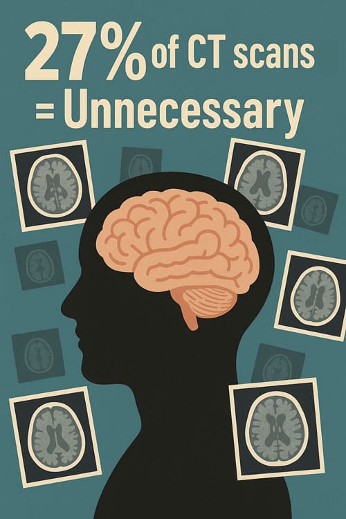
Computed tomography (CT) has become an essential tool in the evaluation of head injuries in emergency departments across the world. Its speed and accuracy in detecting intracranial hemorrhage, skull fractures, and other critical pathology have made it indispensable in acute care. However, concerns about overuse, particularly in mild traumatic brain injury (mTBI), are increasingly recognized. Evidence indicates that approximately 27 percent of CT scans performed for mild head injury are unnecessary when measured against established clinical guidelines. This is troubling given that fewer than 10 percent of adults with mTBI in the United States are ultimately found to have clinically significant brain injuries requiring hospitalization or surgical intervention. The discrepancy between diagnostic yield and actual utilization illustrates the scale of overuse and highlights the need for more consistent, evidence-based decision-making.
Although validated protocols exist, determining when to scan patients with minor head injuries remains a complex clinical challenge. The choice of decision rule can significantly influence imaging rates. Studies suggest that reliance on the Glasgow Coma Scale alone may result in unnecessary scans in up to 37 percent of cases, while the Canadian CT Head Rule reduces this figure to around 30 percent. In pediatric populations, the Pediatric Emergency Care Applied Research Network (PECARN) criteria demonstrate far lower overuse rates, around 10 percent, reflecting the benefits of rigorous and age-specific tools. Yet, even with validated criteria, clinical practice often diverges due to physician discretion, patient expectations, medicolegal pressures, and institutional habits. This variability contributes to inconsistent adherence to guidelines and sustained levels of unnecessary imaging.
The implications of overuse extend beyond diagnostic considerations. At the health system level, the costs are considerable. In Lithuania alone, approximately 1700 patients with minor head trauma undergo head CT annually, generating direct costs of about EUR 121,000. Such expenditures represent a substantial burden on healthcare budgets when viewed alongside the limited diagnostic benefit of many of these scans. From a patient safety perspective, the issue is equally concerning. A non-contrast head CT delivers approximately 2 mSv of ionizing radiation, equivalent to nearly one year of background exposure. While the risk from a single scan is relatively low, repeated or unnecessary imaging increases cumulative exposure and heightens the potential for long-term harm, particularly in younger patients. Overuse also disrupts emergency department workflow, prolongs waiting times, increases strain on radiology services, and often leads to incidental findings that prompt further investigations, additional costs, and potential overtreatment.
Addressing the overuse of CT in mild head injuries requires a multifaceted approach that balances the clinical benefits of imaging against its risks and costs. Strengthening adherence to established guidelines such as the Canadian CT Head Rule and PECARN is a critical step, and embedding these tools into electronic health records or clinical decision support systems can help ensure their consistent application in busy emergency settings. Education for clinicians and trainees remains essential to counter the tendency toward defensive medicine, while patient engagement can help temper demands for unnecessary imaging by clarifying the risks and benefits. Regular audit and feedback mechanisms can also be effective, providing clinicians with data on their imaging patterns and reinforcing evidence-based practice.
Despite progress in defining criteria for appropriate CT use, important knowledge gaps remain. More longitudinal research is needed to better characterize the relationship between mild head trauma, imaging, and long-term outcomes. Comparative studies across different healthcare systems could further refine the applicability of existing tools, while the development of new diagnostic aids, such as serum biomarkers or portable neuroimaging devices, may eventually complement or reduce reliance on CT. Cost-effectiveness analyses are also vital for guiding policy decisions and ensuring that imaging resources are allocated appropriately.
In summary, CT scanning is indispensable in the management of head trauma, but its use in minor injuries is often excessive. Around one-quarter of scans performed for mild head trauma do not meet established criteria, exposing patients to unnecessary radiation, straining healthcare budgets, and slowing emergency department workflows without improving clinical outcomes. The challenge lies in striking the right balance between caution and overuse. By applying validated protocols more consistently, integrating decision support into clinical practice, educating both clinicians and patients, and pursuing further research, healthcare systems can reduce unnecessary imaging while safeguarding patient safety and maintaining high standards of care.
Keywords: head injury, mild traumatic brain injury, CT scan, overuse, clinical guidelines, patient safety, radiation risk
CT Scans in Minor Head Injury: When Are They Used?
Minor head trauma (MHT) represents an enormous diagnostic challenge in emergency settings. Defined as blunt trauma to the head in patients with Glasgow Coma Scale (GCS) scores between 13 to 15, MHT requires careful assessment to identify those at risk for intracranial injuries [1]. Computed tomography (CT) serves as the standard imaging modality for detecting such injuries, though clinical decision rules have been developed to guide its judicious use.
Indications for CT scan head injury in emergency settings
Emergency physicians rely on several established clinical decision rules to determine when a CT scan is warranted following minor head injury. These guidelines aim to identify patients at higher risk while reducing unnecessary radiation exposure and healthcare costs [1].
The Canadian CT Head Rule (CCHR) recommends CT scanning for patients with minor head injury who present with any of these high-risk factors:
- GCS score less than 15 two hours after injury
- Suspected open or depressed skull fracture
- Signs of basal skull fracture
- Two or more episodes of vomiting
- Age 65 years or older
- Amnesia before impact lasting 30 minutes or more
- Dangerous mechanism of injury (pedestrian struck by vehicle, occupant ejected from vehicle, or fall from height of at least 3 feet or five stairs) [2]
Similarly, the New Orleans Criteria (NOC) indicates CT scanning for patients with GCS score of 15 who exhibit headache, vomiting, age older than 60 years, drug or alcohol intoxication, persistent anterograde amnesia, visible trauma above the clavicle, or seizure [2]. The NOC applies only to patients with GCS score of 15, whereas the CCHR may be used for patients with GCS scores from 13 to 15 [3].
The NICE guidelines follow similar principles but include modifications such as delaying CT up to eight hours for “medium risk” patients and classifying coagulopathy as a “high risk” indication requiring immediate CT [3]. Moreover, the guidelines are explicit about pediatric cases, recommending immediate CT scanning for children with post-traumatic seizures, GCS below 14 (or below 15 for infants), suspected skull fractures, or signs of basal skull fracture [1].
CT scanning proves highly effective in detecting acute problems requiring emergent treatment, including hematomas, brain swelling, and skull fractures [4]. Nevertheless, it is noteworthy that CT scans are typically normal in patients with milder forms of traumatic brain injury, including concussion [4]. In fact, more than 75% of treated TBI cases are mild, with only 0.4% to 1% requiring neurosurgical intervention [1].
Role of Glasgow Coma Scale (GCS) in imaging decisions
The Glasgow Coma Scale serves as a fundamental tool for assessing consciousness level and injury severity in head trauma patients. This 15-point scale evaluates a person’s ability to follow directions, move their eyes and limbs, and communicate coherently [5]. Higher scores indicate less severe injuries, with scores of 13-15 typically classified as minor head injury.
GCS scores correlate notably with imaging decision-making. For scores below 13, immediate CT scanning is universally recommended across all clinical decision rules [3]. However, management diverges for patients with GCS scores of 13-15, where additional clinical factors must be considered [2].
Research demonstrates that GCS scores alone may not provide a complete picture of a patient’s prognosis, as they do not give information on potential intracranial lesions visible on imaging studies [6]. Consequently, integration of GCS scores with CT findings generates more precise predictions of outcomes than either method independently [6].
Holly et al. established an association between GCS score-related head injury severity and the risk of accompanying cervical injury, highlighting the scale’s broader utility in trauma assessment [7]. Furthermore, studies show an inverse, approximately linear relationship between mortality and GCS sum score in TBI patients [8].
The ACEP clinical policy demonstrates high sensitivity (92.6%) for identifying pathologies in minor head trauma patients when using appropriate criteria [1]. Nevertheless, the first level of these recommendations alone shows low sensitivity, suggesting that relying solely on primary criteria risks missing many patients with traumatic brain injuries [1].
Understanding Overuse: What Does the Data Say?
Research across multiple healthcare systems reveals a concerning pattern in head imaging practices. The excessive use of CT scans for minor head injuries persists despite established clinical decision rules designed to limit unnecessary radiation exposure.
Pooled overuse rate: 27% across global studies
A comprehensive synthesis of global data demonstrates that approximately 27% of CT scans performed for mild head injuries are unnecessary according to established guidelines [2][9]. This represents more than one in four scans that expose patients to radiation without clinical justification. Certain studies report even higher figures, suggesting that between 10-35% of CT scans for minor traumatic brain injury fail to align with recommended protocols [10].
The extent of overuse varies substantially depending on which clinical decision rule is applied as reference standard. When using the Glasgow Coma Scale (GCS) as the determining factor, the overuse rate reaches 37% [2][9]. Similarly, evaluation against the Canadian CT Head Rule (CCHR) reveals a 30% overuse rate [2][9]. Conversely, pediatric-specific criteria yield lower estimates of unnecessary imaging, with the Pediatric Emergency Care Applied Research Network (PECARN) criterion identifying just 10% of scans as unjustified [2][9].
The National Institute for Health and Clinical Excellence (NICE) guidelines present perhaps the most concerning picture, with 43% of CT scans classified as overuse when measured against these standards [2][9]. The New Orleans Criteria (NOC) yields a more moderate estimate of 18% overuse [2][9]. Yet regardless of which benchmark is applied, the data consistently indicates substantial rates of unnecessary imaging.
Age-related patterns further illuminate this issue. In one notable study, although the overall CT overuse rate was 10.9% for the entire population, this figure jumped dramatically to 37.3% when analyzing only patients younger than 65 years [11]. This suggests younger patients may be particularly vulnerable to excess imaging.
Variation by region: Europe vs Asia vs America
Imaging practices differ markedly across geographic regions. European healthcare systems demonstrate the highest rate of CT overuse in mild head injury patients at 36% [2][9]. Asian healthcare settings follow with 27% overuse [2][9], while American institutions show the lowest rate at 20% [2][9]. These regional variations likely reflect differences in healthcare system structures, medicolegal environments, and implementation of clinical decision rules.
Despite having one of the most thoroughly validated clinical decision rules, the Canadian healthcare system struggled with implementation of the CCHR. Initially, a cluster-randomized trial attempting to reduce CT use through CCHR implementation paradoxically saw imaging rates increase from 63-68% before implementation to 74-76% afterward [12]. The researchers noted this represented an implementation failure rather than a flaw in the rule itself.
In the United States, CT use for pediatric head trauma remained stable at 32% between 2007 and 2015 [7], indicating persistent overuse despite growing awareness of radiation risks. Importantly, CT use showed noteworthy associations with demographic and hospital factors beyond clinical indicators. Children aged ≥2 years were 1.51 times more likely to receive CT scans, while white patients had 1.43 times higher odds of undergoing imaging [7]. Hospital characteristics also played a role, with nonteaching (1.47 times higher odds) and nonpediatric (1.53 times higher odds) facilities showing greater propensity for CT utilization [7].
The pattern of overuse persists regardless of which decision rule serves as the benchmark. Even under optimal implementation, adherence to the CCHR could potentially decrease unnecessary CT scans by approximately 35% [12]. Given that fewer than 10% of mild head injury patients have clinically significant findings on CT, with only 1.3% showing clinically significant changes in some studies [8], the rate of unnecessary scanning represents a substantial opportunity for improvement in global healthcare resource utilization and patient safety.
Decision Rules and Their Impact on CT Utilization
Clinical decision rules profoundly shape CT scan utilization in head injury cases, yet studies demonstrate persistent gaps between guideline recommendations and actual practice. These structured tools aim to identify patients at risk for clinically remarkable brain injuries while reducing unnecessary radiation exposure.
Canadian CT Head Rule (CCHR) overuse rate: 30%
The Canadian CT Head Rule stands as one of the most extensively validated guidelines, designed for patients with minor head injury and Glasgow Coma Scale scores between 13-15. Nonetheless, studies reveal a 30% overuse rate when this rule serves as the reference standard [6]. This means nearly one-third of CT scans performed could potentially be avoided through strict adherence to the CCHR.
Implementation challenges partly explain this overuse phenomenon. In one notable implementation trial, CT imaging rates actually increased from 63-68% before implementation to 74-76% afterward [7]. Researchers attributed this unexpected outcome to implementation failure rather than rule performance. Importantly, the CCHR itself demonstrates excellent sensitivity (100%) for predicting neurosurgical intervention needs while maintaining reasonable specificity (76.3%) [1].
On the positive side, some healthcare systems have successfully reduced CT utilization through CCHR implementation. One multi-center study across 13 Southern California emergency departments noted a 5% absolute reduction in head CT imaging following a CCHR-based intervention [7]. Theoretically, complete adherence to this rule could decrease unnecessary scanning by approximately 35% [7].
NICE guidelines CT scan head injury rate: 43%
Among established protocols, the National Institute for Health and Care Excellence (NICE) guidelines show the highest overuse rate at 43% [6]. These guidelines, widely used throughout the United Kingdom and beyond, were largely derived from the CCHR with certain modifications to improve sensitivity.
The NICE guidelines perform differently depending on the timing of patient presentation. For individuals presenting within 24 hours of injury, sensitivity reaches 98% for detecting serious intracranial injuries. In contrast, this drops to 70% for patients presenting after the 24-hour mark [13]. This temporal difference represents a critical consideration for clinicians applying these criteria.
Cost-effectiveness analysis comparing multiple decision rules identified the CCHR as the most economically sound option [13]. Yet, the NICE guidelines persist as standard practice in many settings due to their institutional integration and familiarity among practitioners.
New Orleans Criteria (NOC) and PECARN comparison
The New Orleans Criteria (NOC) offers an alternative approach, with studies showing an overuse rate of 18% [6]—substantially lower than both CCHR and NICE guidelines. The NOC demonstrates perfect sensitivity (100%) for predicting neurosurgical intervention but suffers from limited specificity (12.1%), resulting in higher CT utilization [1]. If applied strictly, the NOC would mandate CT scanning for 88% of minor head injury patients, versus 52.1% with the CCHR [1].
For pediatric patients, the Pediatric Emergency Care Applied Research Network (PECARN) criteria shows the lowest overuse rate at just 10% [6]. The Canadian Assessment of Tomography for Childhood Head injury (CATCH2) rule presents another pediatric-specific option, with both tools remarkably reducing CT scan usage in children.
Head-to-head comparisons reveal important differences. The CATCH2 rule demonstrates higher sensitivity (100%) and negative predictive value (100%) than PECARN (80% and 98.58%, respectively) [14]. Conversely, PECARN offers better specificity, potentially reducing radiation exposure for approximately 34 more patients per study cohort—at the cost of potentially missing three patients with clinically important traumatic brain injury [14].
Ultimately, each decision rule represents a tradeoff between sensitivity and specificity. Clinical implementation depends not only on statistical performance but likewise on local practice patterns, ease of application, and the relative value placed on detecting every injury versus minimizing unnecessary radiation exposure.
Age and Overuse: Are Younger Patients Scanned More?
Age emerges as a pivotal factor in CT scan utilization for head injuries, with mounting evidence showing distinct patterns of overuse across different age groups. The relationship between patient age and imaging decisions reveals concerning trends that merit close examination by clinicians.
CT overuse in patients under 40: 36%
Patients aged 40 years and below face substantially higher rates of unnecessary imaging compared to their older counterparts. Studies reveal that individuals in this younger age bracket experience CT overuse at a rate of 36% [5]. This means more than one-third of head CT scans performed on younger patients fail to align with established clinical guidelines.
The implications of this overuse pattern are particularly troubling because younger patients face greater lifetime cancer risks from radiation exposure. For children specifically, the stakes are even higher. When comparing age-related radiation risks, infants exhibit a more than ten-fold greater risk than middle-aged adults, primarily due to their increased radiation sensitivity, smaller size, and longer life expectancy [3].
Even within pediatric populations, age-related differences emerge starkly. Analysis of different age groups showed that the chance for undergoing cranial CT increased 14-fold for patients aged 6 to 11 years and 29-fold in children older than 11 years [4]. This escalating pattern suggests that as children approach adolescence, clinical decision-making shifts toward more aggressive imaging strategies.
The setting of care plays a crucial role as well. Children with minor traumatic brain injuries treated in non-pediatric departments undergo neuroimaging (CT or MRI) at a rate of 20.4%, compared to just 5.6% in pediatric departments [15]. Specifically, the rate of cranial CT was 3.8% in pediatric departments versus 18.5% in non-pediatric departments [15]. This translates to a 3.2-fold increased likelihood of CT scanning when pediatric patients are treated in non-pediatric settings [15].
CT overuse in patients over 40: 20%
In contrast, patients over 40 years of age experience a lower but still substantial CT overuse rate of 20% [5]. Several factors may explain this reduced rate of unnecessary imaging. Older patients often present with different symptoms or comorbidities that influence clinical decision-making [5]. Furthermore, clinicians may exercise greater caution when ordering CTs for older patients due to existing health conditions or medications that could increase risks associated with imaging procedures [5].
Yet this lower rate of overuse should not mask a crucial public health reality: because of the sheer volume of CT scans performed on adults, this population accounts for the majority of radiation-induced cancers. Approximately 93,000 of the projected 103,000 radiation-induced cancers from CT examinations (or 91%) will occur in adults despite higher per-scan risks in children [16].
Notably, gender compounds these age-related risks. According to the BEIR-VII lifetime attributable risk assessment, for exposure to a 0.1 Gy radiation dose, females aged 20 years face a 68% greater risk for all cancers than males of the same age [10]. This gender disparity persists across the lifespan, with females maintaining a 20% greater cancer risk even at 60-70 years of age [10].
Ultimately, while younger patients experience higher rates of overuse and greater per-scan risk, the cumulative public health impact remains heavily weighted toward adults due to frequency of scanning. An estimated 61,510,000 patients underwent 93,000,000 CT examinations in 2023, including 2,570,000 (4.2%) children and 58,940,000 (95.8%) adults [16]. This volume translated to approximately 103,000 projected radiation-induced cancers, affecting both pediatric and adult populations [16].
Clinical Settings and Physician Behavior
Physician specialty and practice environment profoundly influence CT scan ordering patterns in head injury cases. Research indicates substantial variation in imaging decisions based on provider type, training, and perceived medicolegal risks.
Emergency medicine vs non-EM physician ordering patterns
The practice setting and provider specialty create notable differences in CT utilization for head injuries. Studies examining Medicare emergency department encounters over 16 years revealed that non-physician providers (NPPs) ordered 5.3% more imaging studies than physicians [11]. This increased ordering manifested across all modalities, with particularly higher rates for CT (7.3%), radiography (3.2%), and other combined modalities (14.2%) [11].
Provider type appears to influence CT utilization across medical specialties. Throughout 2001-2008, the number of emergency department visits involving CT scans managed solely by non-physician providers increased more than sixfold, from 100,626 to 620,296 [2]. Simultaneously, the proportion of emergency CT imaging managed independently by non-physicians grew from 1.5% to 3.6% [2].
Interestingly, when controlling for hospital-level and patient-level variables, visits managed solely by non-physician providers had 0.38 times the odds of CT utilization compared to physician-managed visits [2]. Hence, despite increasing involvement in ordering, non-physicians appear more conservative in their overall CT utilization rates.
Emergency department volume also affects imaging decisions. One study examining the relationship between CT utilization and patient volumes found no statistically measurable difference in CT ordering rates between highest-volume and lowest-volume days [17]. Yet, the rate of negative CT scans increased by 17% during high-volume days [17], indicating that busy clinical environments may impact diagnostic precision without changing overall ordering frequency.
Impact of defensive medicine on CT scan decisions
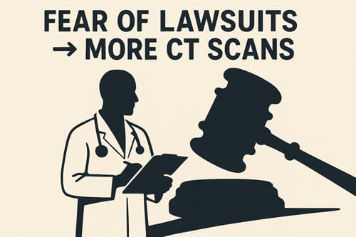
Defensive medicine—the practice of ordering tests primarily to reduce perceived litigation risk rather than for clinical necessity—substantially drives CT overuse in head injury cases. One in three CT scans performed for minor head injuries lacks medical justification [7]. Indeed, fear of missing a diagnosis ranks among the primary factors influencing clinicians’ test-ordering decisions [8].
This defensive approach manifests in various ways. Among emergency physicians, more than 63% acknowledge ordering imaging studies that were not clinically indicated specifically to reduce litigation risks [18]. Risk-averse physicians order substantially more head CT scans than their risk-tolerant colleagues, with one study showing risk-averse emergency physicians ordered head CTs in 56.96% of minor head injury cases versus just 37.37% by risk-tolerant physicians [18].
The doctor-patient relationship emerges as a crucial factor in addressing defensive scanning practices. Research identifies several themes that influence CT decisions: patient engagement, listening, reassurance, identifying patient concerns, and managing patient anxiety [7]. When physicians take time to establish trust through attentive care, patients typically demonstrate greater willingness to accept recommendations regardless of whether CT scanning is indicated [7].
Environmental and systemic factors likewise contribute to defensive scanning. Time pressures, competing interests, distractions, and patient expectations all influence clinical decision-making [8]. Throughout various healthcare systems, both doctors and patients typically overestimate the benefits while underestimating the potential harms of medical interventions, including imaging [8]. Physicians practicing in settings with high malpractice concerns order CT scans at considerably higher rates, with studies showing CT imaging rates are lowest in states that have passed medical liability reform laws [19].
Risks of Over-Scanning: Radiation and False Positives
Beyond the financial implications of unnecessary imaging, over-scanning exposes patients to tangible health risks through radiation exposure and false positive results. The hidden costs of these widespread practices merit careful consideration by clinicians ordering head CT scans.
Radiation exposure from non-contrast CT: 2 mSv
A typical head CT delivers approximately 1 to 2 millisieverts (mSv) of radiation, equivalent to roughly six to eight months of natural background radiation exposure [20]. For context, this radiation dose equals that of approximately 20 standard chest X-rays [9]. Even more concerning, the radiation from a single brain CT scan equals the natural background radiation absorbed over a 3-month period in the United States [12].
While individual risk remains low, the cumulative impact across populations is substantial. Routine overuse of CT imaging contributes to more than 103,000 future cancer cases nationwide [20]. This risk compounds with repeated exposure—patients receiving five or more CT scans show a 2.7% to 12% increase in lifetime cancer risk above baseline [21].
Children face especially troubling risks from ct scan head injury evaluation. Their brain tissue demonstrates greater sensitivity to ionizing radiation, coupled with longer life expectancies that allow more time for radiation effects to manifest [22]. Subsequently, the radiation doses associated with head CT scans may potentially lead to brain tumors later in life, particularly in younger children [23].
The radiation concerns have prompted some institutions to implement low-dose protocols [23]. In Japan, for instance, medical facilities actively work to maintain radiation doses below established diagnostic reference levels, which for non-contrast head CT examinations stand at 85 mGy CTDIvol and 1350 mGy·cm DLP [24].
False positives and unnecessary follow-ups
Equally important, false positive findings and interpretative discrepancies create additional problems. The rate of discordance between general radiologists and neuroradiologists in emergency head CT interpretation reaches 2% overall [25]. More alarmingly, this discordance jumps to 23% for scans where neuroradiologists identified a positive finding [25].
In practice, most ct scans for head injury merely confirm what clinicians already suspect—no remarkable injury exists [20]. One study revealed that 88% of CT scans performed for head trauma were reported as normal without any specific abnormalities [12]. Above all, considering that less than 1% of minor head trauma cases ultimately require surgical intervention, performing head CT scans on all such patients proves unreasonable both economically and from a patient safety perspective [26].
The cascade of unnecessary follow-ups stemming from ambiguous or false positive results can lead to additional testing, specialist referrals, and even invasive procedures. Furthermore, overreliance on imaging sometimes creates a false sense of security that may delay proper diagnosis and treatment for patients who genuinely require intervention [27].
Granted, properly indicated CT scans provide crucial diagnostic information, yet the criteria for ct scan head injury must be carefully applied to balance necessary detection against unnecessary exposure.
Barriers to Reducing Overuse in Practice
Despite established clinical decision rules, several persistent barriers hamper reduction of unnecessary CT scans for head injuries in clinical settings. These obstacles primarily stem from systemic factors rather than knowledge deficits alone, creating a complex challenge for healthcare systems worldwide.
Lack of adherence to clinical decision rules
Implementation of validated guidelines like the Canadian CT Head Rule (CCHR) remains suboptimal in clinical practice. Studies retrospectively analyzing CCHR application reveal that 35.3% to 61.8% of CT scans ordered for minor head injuries failed to meet the rule’s criteria [28]. Concurrently, a separate study found physician non-compliance with the rule reached 28.7% when applied to all adult patients with minor head injuries [28].
Awareness and utilization of these rules varies dramatically by region. An international survey revealed CCHR awareness was highest in Canada at 86% but plummeted to 31% in the United States, while actual usage rates stood at 57% and 12% respectively [28]. Interestingly, provider specialty influences guideline adherence; neurologists order the most non-indicated head CTs while surgeons demonstrate the greatest compliance [28].
Several key barriers impede CCHR implementation according to cognitive task analyzes and provider interviews: memory limitations, attention constraints, decision-making processes, environmental context issues, and social influences [28]. Even with clinical decision support (CDS) systems, adherence to evidence-based guidelines for imaging patients with mild traumatic brain injury reached only 74% in one study—a substantial improvement from 50% baseline but still leaving one in four scans unjustified [29].
Patient expectations and litigation fears
The specter of malpractice litigation fundamentally alters clinical decision-making. In essence, defensive medicine—ordering tests primarily to reduce perceived liability rather than for clinical necessity—drives substantial CT overuse [30]. Physicians frequently cite fear of missing low-probability diagnoses and avoiding malpractice issues as principal contributors to unnecessary imaging [6].
This defensive posture shifts clinical focus from patient wellbeing to legal self-protection, with 78% of physicians believing fear of lawsuits leads to unnecessary testing [30]. Ultimately, this practice adds an estimated $15 billion annually to nationwide healthcare costs [30].
Patient engagement emerges as a crucial counterbalance to defensive imaging practices. Studies identify several themes that influence CT decisions: establishing trust, providing reassurance, identifying patient concerns, and managing anxiety [7]. Physicians who take time to listen and demonstrate care typically find patients more receptive to recommendations regardless of whether imaging is indicated [7].
Research examining fear of malpractice shows mixed results regarding its direct influence on CT ordering. In pediatric head trauma cases, high malpractice fear was associated with increased CT ordering in only one of three case scenarios studied [31]. Nevertheless, states that have undergone tort reform demonstrate measurably lower head CT rates [32].
Strategies to Reduce Unnecessary CT Scans
Effective strategies to combat CT overutilization in head injuries require a multifaceted approach. Evidence-based interventions can substantially reduce unnecessary radiation exposure while maintaining diagnostic accuracy in the emergency setting.
Implementing decision support tools in EMRs
Clinical Decision Support Systems (CDSS) integrated within Electronic Medical Records fundamentally transform imaging appropriateness. These digital mechanisms provide evidence-based guidance at the point of care, ensuring patients receive optimal imaging while avoiding unnecessary procedures [33]. A comprehensive CDSS named CareSelect® Imaging embeds validated criteria directly into the clinical workflow, helping providers identify unnecessary diagnostic imaging through evidence-based guidelines such as the ACR Appropriateness Criteria [34]. One investigation of point-of-care decision support tools demonstrated an 8.2% overall reduction in advanced imaging volume and a striking 61% decrease in duplicate CT and MRI imaging [35]. This implementation not only reduced radiation exposure by 0.27 mSv per patient but also decreased carbon emissions by 13.5% [35].
Training programs on NICE and CCHR guidelines
Firstly, educational interventions prove vital in promoting guideline adherence. Effective training incorporates multiple modalities—including lectures, workshops, and one-on-one discussions—alongside clinical pathway implementation [13]. In practice, this approach might involve sharing audit findings with emergency department teams, adopting guidelines as exclusive reference standards for traumatic brain injury cases, and introducing checklist-style request forms aligned with updated guidance [36]. To maintain knowledge retention, periodic education through a combination of scientific programs and workshops ensures continued reduction in unnecessary imaging [13]. Visual aids such as posters and handouts in clinical areas further reinforce guideline application [13].
Audit and feedback mechanisms for CT ordering
Audit and feedback (A&F) interventions represent a powerful strategy for optimizing CT utilization. Through regular performance summaries, A&F helps clinicians align their practices with evidence-based standards [37]. In a three-arm randomized controlled trial, digital-only feedback reduced CT overutilization by 14%, while hybrid face-to-face/digital approaches achieved an 11% reduction [38]. This improvement translated to 77 fewer cervical spine CT studies monthly, potentially preventing 1,386 unnecessary scans annually if implemented broadly [38]. Correspondingly, meta-analysis reveals A&F interventions result in 1.5 fewer imaging orders per 1,000 patients compared to control interventions [37]. Typically, digital feedback proves more practical than in-person methods due to easier implementation and broader reach [39].

Conclusion 
The overuse of CT scans in minor head injury cases presents a substantial challenge for healthcare systems worldwide. Despite well-established clinical decision rules, approximately 27% of CT scans performed for mild head injuries remain unnecessary. These statistics demand careful consideration, especially given the minimal proportion of patients—fewer than 10% of mild TBI cases—who genuinely require intervention.
Geographic disparities further highlight systemic issues in clinical practice. European healthcare settings demonstrate the highest overuse at 36%, followed by Asia at 27%, while American institutions maintain a comparatively lower rate of 20%. Still, these numbers underscore a universal struggle with balancing thorough evaluation against judicious resource utilization. Though different clinical decision rules yield varying results, each confirms persistent patterns of excessive imaging that transcend regional boundaries.
Age-related patterns reveal another troubling aspect of current practice. Younger patients under 40 years face a disproportionate 36% rate of unnecessary scanning compared to 20% for those over 40. This disparity becomes particularly concerning when considering radiation sensitivity among pediatric populations, who experience exponentially greater lifetime cancer risks from radiation exposure.
Barriers to improvement remain deeply entrenched. The lack of adherence to validated guidelines stems not merely from knowledge gaps but from complex interactions between clinical environments, patient expectations, and medicolegal pressures. Fear of litigation drives defensive medicine practices, with more than 63% of emergency physicians acknowledging they order clinically unnecessary studies primarily to mitigate legal risks.
Nonetheless, effective strategies offer promising paths forward. Clinical decision support systems integrated within electronic medical records have demonstrated an 8.2% reduction in advanced imaging volume and a remarkable 61% decrease in duplicate imaging. Additionally, comprehensive training programs focusing on established guidelines like CCHR and NICE, coupled with regular audit and feedback mechanisms, have yielded measurable improvements in appropriate CT utilization.
Therefore, addressing CT overuse requires a multifaceted approach. Healthcare systems must balance diagnostic thoroughness with patient safety while acknowledging the real-world pressures that influence clinical decision-making. Though the challenge remains substantial, evidence-based interventions can help practitioners navigate these complex waters more effectively, ultimately reducing unnecessary radiation exposure without compromising patient care.
Above all, the goal remains clear—to ensure patients receive appropriate care based on sound clinical judgment rather than defensive practices or systemic pressures. Through continued education, technological support, and organizational commitment, healthcare providers can work toward more judicious use of CT imaging in minor head injury cases, thus enhancing both individual patient outcomes and broader public health.
Key Takeaways
CT scan overuse in minor head injuries represents a tremendous healthcare challenge, with approximately 27% of scans being unnecessary according to clinical guidelines. Here are the essential insights for healthcare providers and patients:
- 27% of CT scans for minor head injuries are unnecessary, exposing patients to 2 mSv radiation (equivalent to 6-8 months of natural background exposure) without clinical benefit.
- Younger patients face higher overuse rates at 36% compared to 20% for those over 40, creating greater lifetime cancer risks for the most vulnerable populations.
- Clinical decision rules show varying effectiveness: NICE guidelines have 43% overuse, Canadian CT Head Rule 30%, while pediatric PECARN criteria achieve only 10% overuse.
- Defensive medicine drives excessive scanning, with 63% of emergency physicians ordering unnecessary CTs primarily to reduce litigation fears rather than clinical necessity.
- Digital decision support systems reduce overuse by 8.2% and duplicate imaging by 61%, proving technology can effectively guide appropriate CT utilization.
The path forward requires balancing diagnostic thoroughness with patient safety through evidence-based guidelines, clinical decision support tools, and addressing the systemic pressures that drive defensive medicine practices.
Frequently Asked Questions:
FAQs
Q1. When is a CT scan necessary after a head injury? A CT scan is typically necessary for head injuries when there are high-risk factors present, such as a Glasgow Coma Scale score below 15, suspected skull fracture, multiple episodes of vomiting, or age over 65. However, for mild head injuries, clinical decision rules like the Canadian CT Head Rule can help determine if imaging is truly needed.
Q2. How much radiation exposure comes from a head CT scan? A typical head CT scan delivers approximately 1 to 2 millisieverts (mSv) of radiation. This is equivalent to about 6-8 months of natural background radiation exposure or roughly 20 standard chest X-rays.
Q3. Are younger patients more likely to receive unnecessary CT scans? Yes, studies show that patients under 40 years old have a higher rate of unnecessary CT scans at 36%, compared to 20% for those over 40. This is particularly concerning due to the increased lifetime cancer risk from radiation exposure in younger individuals.
Q4. How effective are clinical decision rules in reducing unnecessary CT scans? Clinical decision rules show varying effectiveness in reducing unnecessary scans. For example, the Canadian CT Head Rule has an overuse rate of 30%, while the NICE guidelines show 43% overuse. The pediatric-specific PECARN criteria perform best with only 10% overuse.
Q5. What strategies can help reduce unnecessary CT scans for head injuries? Effective strategies include implementing decision support tools in electronic medical records, providing training programs on clinical guidelines, and using audit and feedback mechanisms for CT ordering. These approaches have shown marked reductions in unnecessary imaging while maintaining diagnostic accuracy.
References:
[1] – https://jamanetwork.com/journals/jama/fullarticle/201596
[2] – https://ajronline.org/doi/10.2214/AJR.11.8303
[3] – https://pmc.ncbi.nlm.nih.gov/articles/PMC2672242/
[4] – https://journals.lww.com/md-journal/fulltext/2019/07120/frequency_of_neuroimaging_for_pediatric_minor.32.aspx
[5] – https://journals.plos.org/plosone/article?id=10.1371/journal.pone.0293558
[6] – https://pmc.ncbi.nlm.nih.gov/articles/PMC5815889/
[7] – https://news.yale.edu/2015/11/17/doctor-patient-relationship-key-reducing-ct-scan-overuse
[8] – https://pmc.ncbi.nlm.nih.gov/articles/PMC9206095/
[9] – https://www.brainscope.com/blog/reducing-radiation-exposure-for-mild-head-injured-patients
[10] – https://www.sciencedirect.com/science/article/pii/S1326020023022392
[11] – https://jamanetwork.com/journals/jamanetworkopen/fullarticle/2798248
[12] – https://pmc.ncbi.nlm.nih.gov/articles/PMC10749414/
[13] – https://pmc.ncbi.nlm.nih.gov/articles/PMC7797283/
[14] – https://pmc.ncbi.nlm.nih.gov/articles/PMC8517464/
[15] – https://pmc.ncbi.nlm.nih.gov/articles/PMC6641849/
[16] – https://jamanetwork.com/journals/jamainternalmedicine/fullarticle/2832778
[17] – https://www.sciencedirect.com/science/article/pii/S0735675720303156
[18] – https://pmc.ncbi.nlm.nih.gov/articles/PMC7938907/
[19] – https://onlinelibrary.wiley.com/doi/10.1111/acem.12824
[20] – https://www.healthaffairs.org/do/10.1377/forefront.20250721.419754/full/
[21] – https://www.health.harvard.edu/cancer/radiation-risk-from-medical-imaging
[22] – https://www.aafp.org/pubs/afp/collections/choosing-wisely/40.html
[23] – https://injury.research.chop.edu/blog/posts/reducing-ct-use-mild-traumatic-brain-injury
[24] – https://link.springer.com/article/10.1007/s11604-016-0545-3
[25] – https://ajronline.org/doi/10.2214/ajr.180.6.1801727
[26] – https://www.auntminnie.com/clinical-news/ct/article/15618616/study-finds-ct-is-overused-for-minor-head-injuries
[27] – https://pmc.ncbi.nlm.nih.gov/articles/PMC10783716/
[28] – https://pmc.ncbi.nlm.nih.gov/articles/PMC10290497/
[29] – https://www.annemergmed.com/article/S0196-0644(13)00763-4/fulltext
[30] – https://www.researchgate.net/publication/232493952_Fear_of_
malpractice_liability_and_its_role_in_clinical_decision_making
[31] – https://pubmed.ncbi.nlm.nih.gov/21346679/
[32] – https://onlinelibrary.wiley.com/doi/10.1111/acem.12823
[33] – https://pmc.ncbi.nlm.nih.gov/articles/PMC8754686/
[34] – https://www.acr.org/Clinical-Resources/Clinical-Tools-and-Reference/Clinical-Decision-Support
[35] – https://www.auntminnie.com/clinical-news/ct/article/15661639/pointofcare-decision-support-reduces-unnecessary-ct-mri-imaging
[36] – https://pmc.ncbi.nlm.nih.gov/articles/PMC11334224/
[37] – https://pmc.ncbi.nlm.nih.gov/articles/PMC11152319/
[38] – https://qualitysafety-bmj-com.bibliotheek.ehb.be/content/early/2025/04/02/bmjqs-2024-018374
[39] – https://codex.ucsf.edu/news/editors-pick-new-study-shows-feedback-lowers-overuse-ct-imaging

