New Evidence Shows Strong Connection Between Autism Spectrum and Skin Disorders
Please like and subscribe if you enjoyed this video 🙂
Introduction
Atopic dermatitis (AD) affects up to 20% of children worldwide, making it the most prevalent chronic inflammatory skin condition in pediatrics. Emerging research highlights a notable association between AD and neurodevelopmental disorders, particularly autism spectrum disorder (ASD). Notably, children with AD are 2.3 times more likely to exhibit behaviors falling within the highest severity level of the Autism Diagnostic Observation Schedule (ADOS-2).
This link gains further relevance in light of ASD’s rising prevalence, now affecting approximately 1 in 36 children globally. Studies consistently report a higher prevalence of eczema in children with ASD (28.4%) compared to typically developing peers (15.4%). These children often present with altered epidermal function and heightened skin sensitivity, which may contribute to the increased frequency of skin-related symptoms observed in ASD.
Moreover, severe skin involvement in early childhood has been associated with an odds ratio of 2.51 for subsequent ASD diagnosis, suggesting a bidirectional relationship between inflammatory skin conditions and neurodevelopment. The connection between eczema and autism is not coincidental but likely reflects shared underlying mechanisms, including immune dysregulation, genetic predisposition, and environmental influences that affect both skin barrier integrity and brain development.
This intersection has important clinical and economic implications. Children with ASD incur annual medical costs nearly nine times higher than neurotypical children ($9,980 vs. $1,102), underlining the healthcare burden associated with comorbid conditions.
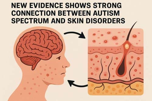
Collectively, these findings underscore the need for integrated care approaches. The skin-brain axis, once considered two separate clinical domains, is increasingly recognized as a key area of overlap between dermatology and neurodevelopment. Early identification and coordinated management of skin conditions in children with or at risk for ASD may offer new opportunities to improve both dermatologic and developmental outcomes.

Shared Epidemiology of Autism and Skin Disorders
Epidemiological investigations consistently demonstrate strong associations between atopic disorders and autism spectrum disorder (ASD), with multiple large-scale studies confirming bidirectional relationships between these conditions. Recent research reveals striking patterns in co-occurrence rates, risk factors, and gender distributions that suggest shared underlying mechanisms.
Prevalence of eczema in children with ASD
The prevalence of eczema among children with ASD varies widely, ranging from 7% to 64.2%, but is consistently higher than in neurotypical populations. One controlled study of 363 individuals with skin diseases reported eczema in 33.9% of those with ASD, compared to 15% of controls. Another study found similar results, with 28.4% of ASD children affected versus 15.4% of controls (adjusted OR = 7.17, 95% CI = 2.56–20.04).
Additional dermatologic findings in ASD populations include:
- Dry skin: 42% in children with ASD vs. 7.2% in controls
- Sensitive skin: 37.6% in ASD vs. >50% with normal skin in controls
Severity of autism appears to influence the rate of atopic conditions. In one study, 80% of children with severe autism (based on Childhood Autism Rating Scale scores) had atopic diseases, compared to 33% in mild-to-moderate ASD and 10% in neurotypical children, suggesting a dose-dependent relationship.
Increased risk of atopic dermatitis in ASD populations
The relationship between ASD and eczema is not only strong but bidirectional. Children with an autistic sibling show increased risk of developing atopic dermatitis (RR = 1.22, 95% CI = 1.09–1.36), with incidence rates of 9.9% vs. 8.1% in controls.
Conversely, having atopic dermatitis greatly raises the likelihood of an ASD diagnosis:
- In China, ASD prevalence among children with atopic dermatitis was 0.99% vs. 0.09% in controls (HR = 8.90, 95% CI = 4.98–15.92)
- In the U.S., ASD prevalence in children with atopic dermatitis was 1.9% vs. 0.9% in non-atopic children (aOR = 1.52)
Importantly, only severe atopic dermatitis was significantly associated with increased ASD risk (OR = 2.54, 95% CI = 1.54–4.21), while mild and moderate cases were not.
Moreover, ASD risk appears to increase with atopic burden. In a cohort of 14,812 individuals with atopic conditions:
- 12.2% had one condition
- 37.2% had two
- 33.4% had three
- 17.1% had four
Data showed a direct correlation between the number of atopic comorbidities and elevated ASD risk.
Gender-based differences in prevalence rates
Gender disparities exist in both autism and skin disorders, though with differing patterns. ASD demonstrates a well-established male predominance, occurring 2-3 times more frequently in males than females. This gender imbalance may be partially attributed to under-recognition in females, who often receive diagnoses at older ages than their male counterparts.
For atopic dermatitis, the gender distribution varies by age. Among children under four years, studies indicate slightly higher prevalence in boys compared to girls (8.7% vs. 5.6%). However, this pattern reverses in adulthood, with higher rates observed in women. Adult prevalence data show rates of 5.7% in men versus 8.1% in women in Japan, and 6.04% in men versus 8.01% in women across Europe and the United States.
The interaction between gender and allergic manifestations in ASD populations reveals additional complexities. One study found allergies (including respiratory allergy and atopic dermatitis) in 57.7% of children with autism, but this figure rose to 80% when considering only females with autism. This suggests that while ASD is less common in females, those females who do have ASD may exhibit higher rates of comorbid atopic conditions compared to males with ASD.
Consequently, these epidemiological connections indicate potential shared biological pathways between autism skin conditions and neurodevelopmental processes, supporting the emerging concept of a skin-brain axis that warrants further investigation for both diagnostic and therapeutic applications.

Environmental and Prenatal Risk Factors
Recent research identifies several prenatal factors that increase risk for both autism spectrum disorder (ASD) and atopic dermatitis (AD), suggesting shared developmental pathways between the brain and skin. Studies increasingly point to critical environmental exposures during gestation that may contribute to the co-occurrence of autism itchy skin manifestations and neurodevelopmental challenges.
Maternal immune activation and cytokine exposure
Maternal immune activation (MIA) during pregnancy represents a major risk factor for autism and skin disorders. Also, maternal asthma stands out as the most common immune condition reported in mothers of children with ASD. The presence of maternal immune conditions is more prevalent in mothers of male children with ASD (31%) compared to female children (18%). Specifically, asthma occurs twice as frequently in mothers of male children with ASD than in mothers of female children with ASD.
MIA operates through several mechanisms that affect fetal development. During maternal immune dysregulation, cytokines and chemokines can cross the blood-brain barrier and placenta, potentially disrupting fetal programming. Indeed, many cytokines do not necessarily need to cross these barriers to exert effects; they can bind receptors at the placental interface, creating downstream impacts on the placenta and fetus.
The inflammatory profile in MIA includes elevated levels of pro-inflammatory cytokines, including IL-4, IL-6, TNF-alpha, and IL-17A. Remarkably, these same inflammatory markers are elevated in both autism and skin disorders like atopic dermatitis and psoriasis. Research demonstrates that IL-6 plays a critical role in promoting MIA-induced ASD-like deficits, and can cross the placenta to directly influence the developing fetus.
Impact of prenatal exposure to phthalates and pollutants
Environmental toxicants represent another shared risk pathway for autism and skin disorders. Prenatal exposure to phthalates has been linked to increased ASD symptoms in children. Specifically, higher maternal concentrations of monoethyl phthalate (mEP) are associated with increased ASD symptoms in boys. Another study found that elevated prenatal di-(2-ethylhexyl) phthalate (DEHP) levels correlated with increased ASD symptoms at ages 2 and 4.
Essentially, phthalates function as endocrine disruptors that can interfere with hormone production and homeostasis. These compounds exert an adjuvant effect on allergen-related immunoglobulin production, enhancing allergic responses through the release of IL-4 from Th2 cells. This mechanism may explain their dual impact on neurodevelopment and skin barrier function.
Alongside phthalates, other environmental exposures like particulate matter, traffic-related air pollution, and heavy metals contribute to immune dysregulation favoring allergic reactions. A meta-analysis revealed that maternal exposure to air pollution was associated with a 1.4-fold increase in ASD risk. Likewise, heavy metals like cadmium and cesium during pregnancy are linked to a 1.8-fold increase in autism risk.
Vitamin D deficiency and seasonal variation in AD/ASD onset
Vitamin D status during pregnancy emerges as another crucial factor connecting autism skin conditions and neurodevelopmental outcomes. Multiple studies report lower serum 25(OH)D concentration in both AD and ASD populations compared to healthy controls. Maternal vitamin D deficiency during pregnancy increases the risk of both conditions in offspring.
Indeed, epidemiological surveys show higher ASD prevalence in urban areas, at higher latitudes, and in locations with more air pollution—all factors that correlate with reduced vitamin D synthesis. Conception during fall in the northern hemisphere is associated with a 6% higher risk of autism compared to summer births. Additionally, being a child of dark-skinned immigrants in colder countries increases the likelihood of developing ASD.
Vitamin D plays dual roles in these conditions. For skin health, it maintains barrier function and reduces inflammation. In neurodevelopment, vitamin D influences myelination and functions as a neuroactive steroid affecting brain development. This dual functionality helps explain the seasonal variations observed in both conditions, with AD often worsening during winter months when vitamin D levels typically decline.

Severity Correlation Between Autism and Eczema
Research examining the relationship between autism spectrum disorder (ASD) and eczema reveals a compelling pattern: children with these comorbid conditions often present with more pronounced symptoms. Beyond mere co-occurrence, evidence points to a relationship between symptom intensity in both conditions, suggesting potential shared biological mechanisms rather than coincidental overlap.
ADOS-2 scores in children with eczema
The Autism Diagnostic Observation Schedule, Second Edition (ADOS-2) represents the gold standard for quantifying autism symptom severity. Recent studies demonstrate that children with both ASD and atopic conditions display higher mean ADOS Calibrated Severity Scores (CSS) on the total scale (7.79±1.51) compared to children without atopic comorbidities (7.16±1.86). This difference, albeit subtle, reflects measurable clinical distinctions between these populations.
Most strikingly, children with atopic conditions were 2.3-2.4 times more likely to receive scores in the ADOS-2 highest severity bracket compared to non-atopic peers. Those specifically reporting comorbid eczema demonstrated even greater severity disparities, with mean ADOS CSS total scores of 7.95±1.62 versus 5.71±1.27 in non-atopic children. Hence, the presence of autism itchy skin manifestations appears to correlate with intensified behavioral presentations across multiple domains.
Interestingly, comparisons between different atopic conditions reveal that children with eczema display more pronounced symptoms than those with asthma or allergies alone. This finding underscores the distinctive relationship between autism and skin disorders, suggesting that skin-brain connections may exceed those of other atopic conditions.
Eczema severity and frequency of clinical visits
The relationship between autism and eczema follows a dose-dependent pattern. Yaghmaie et al. found a direct correlation between parent-reported eczema severity and the prevalence of mental disorders, including autism. Accordingly, a longitudinal study by Liao et al. assessed eczema severity based on frequency of clinical visits, discovering that ASD risk increased proportionally with the number of dermatology appointments.
Perhaps most tellingly, research indicates that only children with severe atopic dermatitis show substantial association with ASD risk (OR=2.54, 95% CI=1.54–42.1), whereas mild (OR=1.07) and moderate cases (OR=0.96) demonstrate no statistically meaningful correlation. This gradient effect provides compelling evidence that inflammatory burden, rather than diagnostic category alone, may influence neurodevelopmental outcomes.
Association with social affect and repetitive behaviors
Domain-specific analyzes reveal that atopic conditions disproportionately impact social functioning in children with ASD. Children with atopic diseases recorded higher scores on the ADOS CSS-Social Affect (SA) subscale (7.63±1.51) relative to non-atopic peers (7.00±2.07). Moreover, they were 2.7-2.9 times more likely to exhibit social difficulties in the severe range.
In contrast, when examining restricted and repetitive behaviors (RRB), no substantial differences emerged between atopic (7.42±1.94) and non-atopic cohorts (7.45±2.22). This selective impact on social domains offers crucial insights for clinicians, suggesting that inflammatory processes may particularly affect neural circuits governing social cognition and interaction.
Among children with autism skin sensitivity, symptom manifestations often include heightened pruritis and itching. The prevalence of these sensory symptoms was markedly elevated in ASD groups compared to controls. Such findings highlight how skin-related discomfort might exacerbate behavioral challenges through sleep disruption, attention difficulties, and heightened irritability—factors that primarily affect social functioning rather than repetitive behavior patterns.
These correlations between autism and skin rashes underscore the importance of comprehensive assessment and treatment approaches that address both dermatological and neurodevelopmental aspects simultaneously.
Immune Dysregulation and Cytokine Profiles
Emerging immunological research highlights the critical role of immune system abnormalities as a potential mechanistic link between autism spectrum disorder (ASD) and skin conditions. The overlapping inflammatory pathways observed in both conditions suggest common biological processes that may explain their frequent co-occurrence.
Elevated IL-4, IL-6, and TNF-alpha in both conditions
Individuals with ASD display distinct inflammatory profiles characterized by elevated pro-inflammatory cytokines. TNF-alpha levels appear substantially higher in children with ASD under 5 years compared to older children and control groups. This elevation correlates with symptom severity across multiple behavioral domains. TNF-alpha concentrations predict ASD phenotypes with considerable accuracy (area under the curve = 0.74), suggesting its potential utility as a biomarker.
IL-6, traditionally classified as a pro-inflammatory cytokine with dual roles in regenerative and anti-inflammatory activities, shows a trend toward elevation in boys with ASD. This cytokine can impair neuronal cellular adhesion, migration, and synapse development. In cerebellum tissue samples from post-mortem ASD subjects, elevated IL-6 levels contribute to altered excitatory-inhibitory circuit balance.
IL-4 levels also demonstrate meaningful alterations, with higher maternal serum IL-4 associated with increased ASD risk in newborns. Interestingly, the plasma cytokine profile in ASD differs from typical inflammatory conditions—some studies report reduced IL-10 (anti-inflammatory) alongside varied patterns of pro-inflammatory markers.
Role of Th2 and Th17 pathways in skin and brain inflammation
The Th2 immune pathway plays a central role in atopic dermatitis, beginning with allergen uptake by Langerhans cells after penetration through the compromised epithelial barrier. These activated cells migrate to the skin where IL-4 promotes Th2 differentiation. Subsequently, keratinocytes produce chemokines that recruit Th2 cells and eosinophils to the skin.
For autism itchy skin manifestations, this altered immune balance carries neurological implications. Mast cells secrete transforming growth factor-β (TGF-β), which promotes the development of Th17 cells and IL-17 production. Elevated IL-17 has been documented in both the serum and immune cells of children with ASD.
Remarkably, IL-17 serves as a common inflammatory mediator in both psoriasis and ASD. Serum IL-17A levels positively correlate with ASD severity, and inhibition of IL-17A signaling improves ASD symptoms in mouse models. Even more compelling, treatment of psoriasis with Secukinumab (an IL-17A inhibitor) alleviates psychological symptoms of ASD in humans.
Autoantibodies and mast cell activation in ASD
Mast cells function as key mediators in autism and skin disorders through their unique ability to respond to non-IgE stimuli and release mediators “differentially” without the typical degranulation seen in allergic reactions. These cells can be activated by environmental and stress triggers, releasing pro-inflammatory and neurotoxic molecules that contribute to brain inflammation.
Increasing evidence points to high brain expression of pro-inflammatory cytokines and circulating antibodies against brain proteins in ASD. Additionally, children with ASD may present with “allergic-like” problems despite normal serum IgE levels and absence of chronic urticaria, suggesting non-allergic mast cell activation.
This dysregulated mast cell response can lead to release of mitochondrial DNA (mtDNA), which acts as an “innate pathogen” causing autoinflammatory responses. Elevated mtDNA has been confirmed in the serum of children with ASD. Furthermore, maternal immune IgE can lead to vertical transmission of atopic dermatitis in newborns by stimulating fetal mast cells.
The autoimmune component manifests through cerebellar-specific autoantibodies found in children with ASD, which correlate with lower adaptive and cognitive function. Moreover, autoantibodies against FcεRIα have been identified, which can cross-link receptors on mast cells and trigger histamine release without traditional allergen exposure.
Therefore, the bidirectional relationship between autism and skin rashes likely stems from these shared immunological pathways that impact both neural and dermal tissues.
Genetic and Epigenetic Overlap
Genetic research unveils underlying molecular connections that may explain the frequent co-occurrence of autism and skin disorders. Multiple studies highlight shared genetic architecture and regulatory mechanisms affecting both neural development and skin barrier function.
Shared gene variants: STAT6, GATA3, FLG
Signal transducer and activator of transcription 6 (STAT6) represents a critical common signaling pathway supporting the etiological correlation between atopy and neurodevelopmental disorders. STAT6 genetic variants associate with atopic conditions through regulation of Th2 immune responses, yet concurrently, STAT6 expresses in the central nervous system with key roles in neuropsychiatric conditions. Four specific STAT6 SNPs (rs3024975, rs841718, rs167769, and rs703817) demonstrate major associations with eczema herpeticum, with odds ratios ranging from 1.40 to 2.14. A 2-SNP haplotype comprising rs167769 and rs324013 shows particularly strong correlation (24.9% vs 9.2%, P = 5.17×10⁻⁶).
GATA3 mutations appear in both atopic dermatitis and ASD pathogenesis. This transcription factor, essential for T-cell development and differentiation, creates another genetic bridge between these conditions. Filaggrin (FLG) gene mutations constitute the most important known risk factors for atopic dermatitis and represent another potential shared genetic element.
MicroRNA involvement: miR-146a and miR-155
MicroRNAs play fundamental roles in molecular and cellular mechanisms underlying both conditions. miR-146a, upregulated in various neurodevelopmental disorders, demonstrates high expression throughout the cortex, hippocampus, and amygdala—key structures for higher cognitive functioning. In mouse models, abnormal miR-146a expression leads to impaired neuronal dendritic arborization and shriveled dendritic trees, producing the defective neural connectivity typical of ASD.
For skin conditions, increased miR-146a expression appears in lesional skin of AD patients. This microRNA inhibits nuclear factor κB (NF-κB)-mediated proinflammatory cytokines, alleviating inflammation linked to atopic conditions. Regarding miR-155, although its role in ASD remains unclear, this microRNA contributes to T helper type 17 (Th17) cell differentiation in atopic dermatitis.
Epigenetic modulation via environmental triggers
Environmental factors influence gene expression in both conditions through epigenetic mechanisms. DNA methylation, transcriptional regulation, and post-translational changes in histone proteins contribute to the pathogenesis of both disorders. Altered epigenetic regulation via genomic alterations (SNPs or copy number variants) can lead to epigenetic dysregulation ultimately causing ASD.
Environmental exposures—including chemical compounds, heavy metals, nutrition, and maternal stress—affect programming of offspring’s epigenome. Notably, prenatal exposure to endocrine-disrupting chemicals modifies epigenetic markers, potentially influencing both skin and neurological development. Nutritional factors like folate and vitamins B2, B6, and B12 participate in one-carbon metabolism involved in DNA methylation processes that affect both conditions.
Comorbidities and Clinical Implications
Children with autism spectrum disorder (ASD) face a complex array of medical comorbidities that extend beyond neurological symptoms, creating substantial challenges for patients, families, and healthcare providers. These interconnected conditions demand integrated clinical approaches that address multiple body systems simultaneously.
ADHD and allergic rhinitis in ASD patients
The relationship between ASD and attention deficit hyperactivity disorder (ADHD) represents one of the most frequent neurodevelopmental comorbidities. Nearly 20% of individuals with ASD meet ADHD diagnostic criteria, with evidence suggesting these conditions share overlapping biological mechanisms. Patients with concurrent ASD and ADHD diagnoses experience greater illness severity and functional impairment.
In parallel, allergic rhinitis emerges as the most prevalent atopic condition among children with ASD-ADHD comorbidity. These children exhibit markedly higher rates of allergic rhinitis (59.0%) compared to controls (38.8%). The dual diagnosis group demonstrates a greater average number of allergic comorbidities (1.47) versus controls (0.97). Female individuals with ADHD appear particularly susceptible to developing allergic rhinitis compared to their male counterparts.
Sleep disturbances and gastrointestinal symptoms
Sleep disorders affect approximately 80% of children with ASD, manifesting as bedtime resistance, insomnia, parasomnia, and daytime sleepiness. These disruptions correlate directly with behavioral challenges, as children experiencing poor sleep demonstrate increased irritability, oppositionality, and difficulty with attention.
Gastrointestinal (GI) problems represent another pervasive comorbidity, occurring in 46-84% of autistic children. Common manifestations include chronic constipation, diarrhea, gastroesophageal reflux, and abdominal discomfort. These symptoms strongly correlate with autism severity, suggesting children with more pronounced autistic features typically experience more severe GI dysfunction.
Importantly, GI symptoms and sleep problems demonstrate bidirectional relationships. Children with both autism itchy skin conditions and GI symptoms experience higher rates of sleep disturbance (50%) than those without GI issues (37%).
Implications for early screening and diagnosis
The complex interplay between autism skin disorders, sleep problems, and gastrointestinal dysfunction necessitates comprehensive assessment protocols. To that end, clinicians should recognize that behaviors commonly attributed to autism—including irritability, self-injury, and increased repetitive movements—may actually reflect underlying medical conditions.
For instance, facial grimacing, teeth grinding, and excessive chewing often represent expressions of GI discomfort in children with limited verbal communication abilities. Early identification of these comorbidities can substantially improve quality of life while potentially reducing behavioral challenges.
Nevertheless, diagnostic overshadowing remains problematic, as ASD diagnosis sometimes delays recognition of comorbid conditions like anxiety or ADHD. Over time, this comprehensive approach to assessment may reduce healthcare costs, which currently average nine times higher for children with ASD ($9,980) compared to neurotypical peers ($1,102).

Conclusion
Mounting evidence supports a strong, bidirectional relationship between autism spectrum disorder (ASD) and atopic dermatitis (AD). Children with ASD are more likely to experience eczema, while those with severe AD show increased risk of being diagnosed with ASD. This connection is not simply statistical, it reflects complex biological and environmental interactions involving both the skin and brain.
Central to this relationship are overlapping immune pathways. Elevated levels of pro-inflammatory cytokines, particularly IL-4, IL-6, TNF-alpha, and IL-17, are implicated in both neuroinflammation and skin inflammation. These cytokines disrupt neural function and skin barrier integrity, suggesting parallel inflammatory processes in both systems.
Genetic factors also play a key role. Variants in genes such as STAT6, GATA3, and FLG contribute to dysregulation of immune responses and cellular development in both conditions. These shared molecular pathways point to a common biological foundation underlying ASD and AD.
Environmental exposures further reinforce the link. Maternal immune activation, exposure to environmental toxins (e.g., phthalates), and vitamin D deficiency have all been associated with increased risk for both ASD and AD. Notably, there is a dose-response relationship: children with severe AD are at a markedly higher risk of ASD compared to those with mild or moderate forms.
These findings have clear implications for clinical care. Skin discomfort may exacerbate behavioral symptoms in children with ASD and go unrecognized in the absence of dermatologic evaluation. Conversely, children with severe eczema should be screened for developmental concerns, as early identification can improve outcomes.
The economic burden is considerable. Children with ASD already incur significantly higher healthcare costs than neurotypical peers. Undiagnosed or unmanaged skin conditions may further increase this burden by driving up healthcare utilization and affecting quality of life.
Recognizing the skin-brain connection calls for integrated, multidisciplinary care. Pediatricians, dermatologists, neurologists, and mental health professionals must collaborate to develop coordinated assessment and treatment strategies.
Research should now focus on targeted therapies that address shared mechanisms such as anti-inflammatory agents, microbiome-based interventions, and immunomodulatory treatments with the goal of managing both conditions more effectively.
Frequently Asked Questions:
FAQs
Q1. Is there a connection between autism and skin conditions? Yes, recent research shows a strong connection between autism spectrum disorder (ASD) and skin conditions, particularly atopic dermatitis (eczema). Children with ASD have a higher prevalence of eczema compared to typically developing peers, and those with severe eczema show an increased risk of developing ASD.
Q2. How do environmental factors contribute to both autism and skin disorders? Environmental factors like maternal immune activation during pregnancy, exposure to phthalates and pollutants, and vitamin D deficiency have been linked to increased risk of both autism and skin disorders. These factors can affect fetal development and immune system regulation, potentially contributing to both conditions.
Q3. Are there shared genetic factors between autism and skin conditions? Yes, genetic research has identified several shared gene variants, including STAT6, GATA3, and FLG, that contribute to both autism and skin disorders. These genes play roles in immune regulation and cellular development, affecting both neural circuits and skin barrier function.
Q4. How does the severity of eczema relate to autism symptoms? Studies have shown a correlation between eczema severity and autism symptom intensity. Children with both ASD and atopic conditions often display higher scores on autism diagnostic tests, particularly in social affect domains. The relationship appears to be dose-dependent, with more severe eczema associated with increased autism risk and symptom severity.
Q5. What are the clinical implications of the autism-skin disorder connection? The connection between autism and skin disorders has major clinical implications. It suggests the need for comprehensive assessment protocols that address both dermatological and neurological symptoms. Healthcare providers should consider routine dermatological evaluation for children with ASD and neurodevelopmental screening for those with severe atopic conditions to ensure proper diagnosis and treatment of both conditions.
References:
[1] – https://pmc.ncbi.nlm.nih.gov/articles/PMC10932299/
[2] – https://pmc.ncbi.nlm.nih.gov/articles/PMC11432970/
[3] – https://pmc.ncbi.nlm.nih.gov/articles/PMC10491411/
[4] – https://pmc.ncbi.nlm.nih.gov/articles/PMC10619695/
[5] – https://www.sciencedirect.com/science/article/pii/S0925443910002954
[6] – https://pmc.ncbi.nlm.nih.gov/articles/PMC8085719/
[7] – https://pmc.ncbi.nlm.nih.gov/articles/PMC3205328/
[8] – https://pmc.ncbi.nlm.nih.gov/articles/PMC11354676/
[9] – https://www.nature.com/articles/s41398-022-02185-5
[10] – https://www.researchgate.net/publication/363919978_Eczema_and_
related_atopic_diseases_are_associated_with_increased_symptom_
severity_in_children_with_autism_spectrum_disorder
[11] – https://www.autismeye.com/eczema-skin-disorders-autism/
[12] – https://www.psypost.org/eczema-linked-to-increased-symptom-severity-in-children-with-autism-spectrum-disorder/
[13] – https://www.mdpi.com/2077-0383/14/2/469
[14] – https://pmc.ncbi.nlm.nih.gov/articles/PMC5669898/
[15] – https://pmc.ncbi.nlm.nih.gov/articles/PMC9626859/
[16] – https://bmcpediatr.biomedcentral.com/articles/10.1186/
s12887-024-05182-3
[17] – https://www.sciencedirect.com/science/article/pii/S0022202X15425473
[18] – https://www.mdpi.com/1422-0067/25/16/8936
[19] – https://pmc.ncbi.nlm.nih.gov/articles/PMC6352260/
[20] – https://pmc.ncbi.nlm.nih.gov/articles/PMC9222497/
[21] – https://pmc.ncbi.nlm.nih.gov/articles/PMC4881129/
[22] – https://www.sciencedirect.com/science/article/abs/pii/S1750946714001664
[23] – https://pmc.ncbi.nlm.nih.gov/articles/PMC11493030/
[24] – https://pubmed.ncbi.nlm.nih.gov/26384115/
[25] – https://pmc.ncbi.nlm.nih.gov/articles/PMC5683266/
[26] – https://www.frontiersin.org/journals/psychiatry/articles/
10.3389/fpsyt.2023.1265472/full

