Can MRI Detect Old Strokes? New Findings on Silent Brain Infarcts

Introduction
The question of whether an old stroke appears on magnetic resonance imaging (MRI) holds remarkable clinical importance, particularly in the context of silent brain infarcts (SBIs). These lesions, often asymptomatic, are increasingly recognized as markers of underlying cerebrovascular pathology and predictors of future neurological events. Advances in neuroimaging have revealed that SBIs are far more prevalent than previously thought. Population-based studies estimate their occurrence in approximately 8% to 28% of individuals, with even higher rates observed in specific cohorts. For example, data from the Cardiovascular Health Study demonstrated that nearly 28% of participants exhibited radiological evidence of silent infarction despite the absence of overt clinical stroke symptoms.
Although SBIs are termed “silent,” their clinical implications are anything but benign. Individuals with MRI-detected silent infarcts experience a substantially higher risk of subsequent cerebrovascular events. The incidence of clinically apparent stroke in this population has been reported at approximately 18.7 per 1,000 person-years, nearly double the rate observed in individuals without SBIs, whose incidence is about 9.5 per 1,000 person-years. Longitudinal studies have further documented an annual SBI incidence of 2% to 4%, underscoring the ongoing risk of new infarction even in asymptomatic individuals. Beyond stroke risk, SBIs are associated with cognitive decline, gait disturbances, and increased overall morbidity in older adults.
MRI remains the most sensitive modality for detecting both acute and chronic infarcts. The imaging characteristics of old strokes are well-defined: chronic infarcts typically appear hypointense on T1-weighted sequences and hyperintense on T2-weighted and fluid-attenuated inversion recovery (FLAIR) images. These signal changes reflect tissue loss, gliosis, and encephalomalacia secondary to previous ischemic injury. In contrast, acute infarcts often demonstrate restricted diffusion on diffusion-weighted imaging (DWI) and corresponding low apparent diffusion coefficient (ADC) values, which can help distinguish them from chronic lesions. Over time, the diffusion restriction resolves, and chronic infarcts evolve to display the characteristic imaging features of tissue loss and gliotic scarring.
The burden of silent cerebral infarction is particularly pronounced among pediatric patients with sickle cell anemia (SCA). In this population, approximately one in four children develops silent infarcts before the age of six, and roughly one-third before the age of fourteen. These findings highlight the need for regular neuroimaging surveillance and early intervention strategies in high-risk groups. Silent infarcts in children with SCA are linked not only to an elevated risk of overt stroke but also to measurable deficits in cognitive performance and academic achievement.
In summary, old strokes do appear on MRI, and their identification carries critical prognostic value. MRI not only enables the detection of silent and chronic infarcts but also helps distinguish them from acute lesions based on characteristic signal patterns and diffusion properties. The presence of these radiological findings should prompt clinicians to evaluate underlying vascular risk factors, initiate appropriate preventive measures, and monitor for potential neurological or cognitive sequelae. Future research should continue to refine MRI-based biomarkers for infarct aging and investigate strategies to mitigate the long-term impact of silent cerebrovascular disease.
Keywords: magnetic resonance imaging, silent brain infarcts, stroke, sickle cell anemia, neuroimaging, cerebrovascular disease
What Is a Silent Brain Infarct (SBI)?
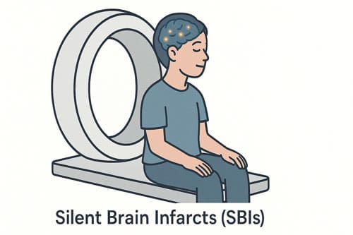
Silent brain infarcts (SBIs) represent a distinct and underrecognized neurological entity that occurs far more frequently than clinically apparent strokes. Although these lesions do not produce the overt neurological deficits typically associated with acute cerebrovascular events, their presence carries important implications for long-term neurological and cognitive health.
Definition and Distinction from Overt Stroke
Silent brain infarcts differ fundamentally from overt strokes in their clinical manifestation. Both conditions result from vascular occlusion leading to localized cerebral tissue damage; however, SBIs are characterized by the absence of acute stroke symptoms or discernible neurological deficits. This asymptomatic nature often renders them undetected without neuroimaging, posing a diagnostic challenge since affected individuals usually remain unaware that a cerebrovascular event has occurred.
By definition, SBIs are asymptomatic brain infarctions identified incidentally through neuroimaging studies, most often during evaluations for unrelated conditions. Magnetic resonance imaging (MRI) serves as the diagnostic modality of choice because of its superior sensitivity and specificity compared to computed tomography (CT). The asymptomatic presentation is often attributed to the relatively small size of the infarcts, yet this explanation alone is insufficient, as similarly small lacunar infarcts can produce overt neurological symptoms. Therefore, the clinical silence of SBIs is likely influenced by both lesion size and anatomical location, particularly when lesions occur in regions with functional redundancy or non-eloquent brain areas.
Clinical Implications and Prognostic Significance
Despite their asymptomatic presentation, SBIs are not clinically benign. Increasing evidence indicates that these lesions are associated with subtle neurological and cognitive impairments, mood and psychiatric disturbances, and a substantially elevated risk of future cerebrovascular events. Multiple population-based studies have shown that the presence of an SBI more than doubles the risk of developing an overt stroke and dementia later in life. Furthermore, individuals with SBIs often exhibit diminished cognitive performance on neuropsychological testing, particularly in domains related to executive function and processing speed. These findings suggest that SBIs may represent an early marker of cerebrovascular pathology and neurodegenerative risk, warranting proactive clinical recognition and management.
MRI-Based Diagnostic Criteria
The diagnosis of SBIs relies on characteristic MRI findings. Typically, an SBI is defined as a focal lesion measuring at least 3 mm in diameter that appears hyperintense on T2-weighted and fluid-attenuated inversion recovery (FLAIR) sequences and demonstrates corresponding hypointensity on T1-weighted images. These imaging features help distinguish SBIs from other white matter abnormalities such as demyelination, gliosis, or dilated perivascular spaces.
Radiological differentiation between SBIs and perivascular (Virchow–Robin) spaces is particularly important. While both can appear as small, rounded lesions in the deep white matter or basal ganglia, perivascular spaces are typically smaller than 3 mm, exhibit linear or ovoid morphology with a perivascular orientation, and maintain CSF-like signal intensity across all MRI sequences. Enlarged perivascular spaces exceeding 3 mm may occur in up to 30 percent of older adults and account for up to 20 percent of SBI misclassifications. Proton-density imaging and high-resolution FLAIR sequences can assist in differentiating these entities, as infarcts typically demonstrate surrounding gliosis and altered tissue signal intensity that perivascular spaces lack.
Typical Locations and Pathophysiology
SBIs display a characteristic distribution pattern within the brain. The basal ganglia are the most frequently affected region, accounting for the majority of SBI cases in several large-scale population studies. In one study of 217 individuals with SBIs, 171 (approximately 79 percent) of lacunar-type infarcts were located in the basal ganglia. Other commonly affected areas include the subcortical and deep white matter, particularly within the frontal lobes, as well as the thalamus. Less frequently, SBIs are observed in the cerebral cortex, cerebellum, and brainstem.
The predilection for subcortical and basal ganglia involvement reflects the underlying vascular pathology. Most SBIs are lacunar in nature, resulting from occlusion of small perforating arteries. Hypertensive small-vessel disease is the predominant etiological mechanism, although other contributors such as diabetes mellitus, dyslipidemia, and endothelial dysfunction play important roles. These lacunar infarcts typically measure between 3 and 15 mm in diameter and often exhibit wedge-shaped, ovoid, or irregular morphologies. Their imaging characteristics often resemble cerebrospinal fluid signal intensity due to tissue necrosis and cavitation.
However, not all SBIs undergo cavitation. Longitudinal imaging studies suggest that between 30 and 80 percent of infarcts remain non-cavitated, persisting as areas of T2 hyperintensity and T1 hypointensity without the development of a well-defined cavity. This variability complicates radiologic interpretation and may influence the visibility of older lesions on MRI, particularly when assessing chronic infarctions.
Silent brain infarcts represent a clinically notable but often overlooked marker of cerebrovascular disease. Although asymptomatic, these lesions indicate underlying small-vessel pathology and confer an increased risk of future stroke, cognitive decline, and dementia. Accurate identification through MRI and differentiation from other white matter changes are essential for appropriate diagnosis and risk stratification. Given their prognostic implications, recognition of SBIs should prompt comprehensive vascular risk assessment and management aimed at preventing further cerebrovascular injury.
How MRI Detects Old Strokes
Magnetic resonance imaging (MRI) remains the most sensitive neuroimaging method for detecting old strokes, offering distinct advantages over computed tomography in visualizing both acute and chronic cerebral infarctions. The ability to detect old strokes through MRI depends on understanding the evolution of signal characteristics across various sequences.
T1 and T2 signal changes in chronic infarcts
Chronic infarcts display characteristic signal patterns that evolve predictably over time. Initially, increased tissue water results in prolongation of T1 and T2 relaxation times. T2 changes appear after 6-8 hours, whereas T1 changes typically develop more slowly, with only 50% of patients showing T1 alterations at 24 hours [9]. As the infarct evolves into the chronic stage (2 weeks-2 months), several key transformations occur:
- Restoration of the blood-brain barrier
- Resolution of vasogenic edema
- Removal of necrotic tissue
- Development of local brain atrophy and gliosis
These pathological changes manifest radiologically as cavity formation and ex vacuo dilatation of adjacent ventricles [9]. In chronic lesions, T1-weighted images reveal hypointensity while T2-weighted images demonstrate persistent hyperintensity. This combination of signal characteristics allows radiologists to identify old strokes with high specificity.
Cortical laminar necrosis, representing neuronal ischemia with gliosis and layered deposition of fat-laden macrophages, manifests as T1 hyperintensity of the cortex. This feature becomes visible approximately 2 weeks post-infarction and reaches peak visibility between 1-3 months [9]. Contrary to previous assumptions, this T1 shortening does not result from hemorrhage but rather from neuronal necrosis and denatured proteins.
Role of FLAIR and DWI in identifying infarct age
Fluid-attenuated inversion recovery (FLAIR) and diffusion-weighted imaging (DWI) sequences play crucial roles in determining infarct age. FLAIR can detect abnormal signals earlier than other conventional sequences, sometimes revealing changes within 3 hours after stroke onset [2]. Conversely, DWI stands out as the most sensitive sequence for detecting acute infarctions, with sensitivity ranging from 88% to 100% and specificity between 95% and 100% [2].
The relationship between DWI and FLAIR signals provides valuable information about infarct age. A positive signal on DWI without corresponding FLAIR hyperintensity (DWI-FLAIR mismatch) indicates the stroke likely occurred between 4.5 and 6 hours prior to imaging, with 62% sensitivity and 78% specificity [8]. Studies have shown that infarct lesions with DWI-FLAIR mismatch typically undergo scanning earlier (3.8 ± .3 vs. 7.5 ± .3 hours from onset) and tend to be smaller (8.9 ± 2.3 vs. 43.1 ± 11.9 mL) compared to lesions without mismatch [7].
Throughout the evolution of an infarct, apparent diffusion coefficient (ADC) values follow a predictable pattern: decreasing in the first 7-10 days, then pseudo-normalizing, and eventually increasing [8]. This temporal relationship between MRI sequences helps identify patients with “unknown onset” or wake-up strokes.
Can an MRI tell how old a stroke is?
MRI can indeed provide reasonably accurate estimates of stroke age through assessment of multiple sequence parameters. Research demonstrates that visual inspection of FLAIR and DWI images can identify strokes within 3 hours of onset with 94% sensitivity and 97% specificity [10]. Moreover, the FLAIR ratio shows positive correlation with time from symptom onset and can identify patients imaged within 3 hours using a 7% FLAIR ratio cutoff (90% sensitivity, 92% specificity) [10].
For infarct lesions exceeding 15 mL, the DWI-FLAIR mismatch demonstrates 100% specificity and positive predictive value for acute infarcts within 4.5 hours of onset [7]. Consequently, this mismatch has become an important biomarker for timing stroke onset, especially in cases where symptom onset time remains unknown.
Recent advances include machine learning algorithms that analyze multiple imaging features beyond visual inspection. These automated systems have shown superior sensitivity (>70%) with comparable specificity (>85%) to human evaluation in identifying patients within therapeutic time windows [7]. Such technological innovations may soon enhance clinical practice by offering more precise timing of cerebral infarctions.
MRI Features That Differentiate Old vs New Strokes
Distinguishing between acute and chronic cerebral infarcts requires understanding the temporal evolution of stroke on MRI. Radiologists rely on specific imaging biomarkers that change predictably over time to determine whether a lesion represents a new stroke or an old infarct.
DWI hyperintensity for acute infarcts
Diffusion-weighted imaging (DWI) serves as the cornerstone for identifying acute ischemic stroke, with sensitivity reaching 88% to 100% and specificity between 95% to 100% [11]. The underlying mechanism involves restricted Brownian motion of water molecules within ischemic tissue. After arterial occlusion, impaired mitochondrial function leads to ATP depletion, causing Na-K pump malfunction and subsequent cellular swelling. This pathophysiological cascade shifts extracellular water to the intracellular compartment, thereby restricting water molecule diffusion that manifests as hyperintensity on DWI [11].
DWI signal changes develop remarkably quickly—emerging within minutes of vessel occlusion [4]. Subsequently, these changes reach maximum visibility between 1-4 days post-stroke [12]. Throughout the first week, infarcted parenchyma consistently exhibits high DWI signal coupled with low apparent diffusion coefficient (ADC) values [12].
Interestingly, not all DWI-negative patients truly lack infarction. Recent meta-analyzes reveal that approximately 6.8% of acute ischemic stroke cases present as DWI-negative initially [5]. Moreover, in one study, 22.5% of initially DWI-negative patients with clinical stroke symptoms demonstrated delayed lesion appearance on follow-up imaging [5]. This phenomenon termed “DWI-positive conversion” holds prognostic importance, as these patients face higher risks of recurrent stroke and mortality [5].
Atrophy and encephalomalacia in chronic lesions
As strokes age, they undergo characteristic transformations culminating in encephalomalacia—defined as brain tissue softening resulting from liquefactive necrosis [13]. Radiologically, this appears as focal volume loss with CSF-like signal characteristics across multiple MRI sequences [13].
On T1-weighted images, chronic infarcts demonstrate hypointensity, while T2-weighted and FLAIR sequences show hyperintensity [13]. ADC values in these regions become elevated above normal brain tissue—a feature directly opposite to the restricted diffusion seen in acute infarcts [12].
Encephalomalacia from old infarcts typically presents with these distinctive features:
- Focal volume loss exceeding 15mm in diameter [14]
- CSF-like attenuation across multiple MRI sequences [14]
- Ex vacuo ventricular enlargement and widening of cortical gyri [15]
- Surrounding gliosis (proliferation of glial cells) [13]
Importantly, pre-existing non-disabling encephalomalacia has been identified in 24.5% of patients undergoing endovascular thrombectomy in one cohort study [16]. However, contralateral encephalomalacia (relative to a new acute event) and larger encephalomalacic lesions (>15mm) were associated with unfavorable outcomes after intervention [16]. Hence, identifying these chronic changes carries both diagnostic and prognostic value.
Absence of restricted diffusion in old stroke on MRI
Perhaps the most reliable differentiator between new and old strokes involves diffusion characteristics. As infarcts age, ADC values follow a predictable temporal evolution: initially decreasing, then gradually normalizing, and finally increasing above baseline [12]. This “pseudo-normalization” typically occurs between 10-15 days post-infarction [4].
After pseudo-normalization, the ADC continues rising until chronically infarcted tissue demonstrates facilitated (not restricted) diffusion [6]. However, DWI signal patterns during this transition merit careful interpretation. Even after ADC normalization, DWI may remain hyperintense due to “T2 shine through”—persistent T2/FLAIR signal elevation contributing to brightness on DWI despite normal or elevated ADC [4].
Later still, as gliosis develops and encephalomalacic changes progress, DWI signal intensity actually decreases despite T2 hyperintensity—a phenomenon termed “T2 washout”—because the overwhelmingly facilitated diffusion counteracts the T2 effects [12].
Recent studies have also revealed that some infarct regions remain hyperintense on DWI even at one month post-stroke, occurring in 64% of patients in one study [1]. These persistently hyperintense regions occurred predominantly in white matter and showed different perfusion recovery patterns compared to areas that normalized, suggesting heterogeneous tissue responses to ischemia [1].
Common Mimics of Silent Infarcts on MRI
Accurate identification of silent brain infarcts requires careful differentiation from several conditions that can mimic their appearance on MRI. Throughout clinical practice, radiologists encounter various entities that present similar imaging characteristics to infarcts, yet reflect entirely different pathological processes.
Dilated Virchow-Robin spaces
Virchow-Robin spaces represent perivascular spaces that surround small vessels as they extend into brain tissue. These spaces can enlarge under undetermined conditions, creating dilated Virchow-Robin spaces (dVRS) that frequently mimic lacunar infarcts on MRI [17]. The distinction between cavitated infarction and dilated perivascular spaces presents the most important diagnostic difficulty when evaluating potential silent infarcts [18].
Key differentiation features for dVRS include:
- Location along perforating or medullary arteries, or in the lower third of basal ganglia [18]
- Linear, round, or ovoid shape, depending on slice direction [19]
- Absence of a hyperintense rim around suspected lesions on fluid-attenuated inversion recovery (FLAIR) images [18]
- Size typically smaller than 2×1 mm when excluding lesions from lower basal ganglia [20]
Prevalence studies reveal dVRS in 100% of elderly subjects when using high-resolution 3D MRI with voxel size of 1.0×0.98×0.98 mm³, alongside dVRS exceeding 3 mm in approximately one-third of them [17]. The most common location occurs at the proximal part of lenticulostriate arteries, with prevalence twice as high in this area compared to along cortical-medullary arteries [17].
Periventricular leukomalacia
Periventricular leukomalacia (PVL) represents softening of white brain tissue near the ventricles that can be confused with old strokes on imaging. PVL primarily occurs in premature infants whose brain tissues are more fragile, resulting from hypoperfusion of end arteries that supply periventricular white matter [21].
On MRI, PVL manifests as:
- Periventricular zones of increased signal intensity on T2-weighted and inversion recovery images
- Hypointensity on T1-weighted images [21]
- Specific predilection for peritrigonal areas [21]
- Late findings including loss of white matter leading to gliosis and cavitation [21]
PVL represents the second most common hypoxic brain injury in premature infants, accounting for 4-26% of cases, behind only intraventricular and periventricular hemorrhage (30-55%) [21]. Clinical presentations range from spastic diplegia to cortical blindness, alongside cerebral visual impairment that affects a child’s learning ability [21].
Posterior reversible encephalopathy syndrome (PRES)
Posterior reversible encephalopathy syndrome represents a clinico-neuro-radiological diagnosis that can mimic stroke. PRES manifests with clinical features including headache, vomiting, altered sensation, visual disturbances, seizures, and focal deficits [3]. Radiologically, PRES shows predominant posterior leukoencephalopathy with vasogenic edema [3].
While MRI remains superior for PRES diagnosis, computed tomography may be necessary for patients with contraindications to MRI. Notably, only 32% and 74% of patients had contributory CT findings on days 1 and 2 respectively, suggesting value in repeated scanning when MRI is unavailable [3]. The cerebral regions affected by PRES include occipital, parietal, frontal, and temporal lobes in decreasing order of frequency [3].
Given the advent of stroke thrombolysis, patients with PRES risk unnecessary thrombolytic treatment due to normal initial CT findings and stroke-like presentation. Consequently, maintaining high clinical suspicion based on history and examination remains essential for preventing inappropriate interventions and permanent brain damage [3].
Distinguishing these mimics from true silent infarcts proves vital not only for diagnostic accuracy but also for appropriate clinical management and prognostication. For radiologists evaluating whether old strokes appear on MRI, recognizing these common mimics facilitates more precise interpretation of neuroimaging findings.
Prevalence and Risk Factors for Silent Strokes
Silent brain infarcts (SBIs) occur far more commonly than previously recognized, with population-based studies revealing an astonishing prevalence across diverse demographics. Understanding the epidemiology and risk factors associated with these lesions provides key insights into their detection and management.
Age-related increase in SBI prevalence
The likelihood of harboring silent strokes rises dramatically with advancing age. In the landmark Rotterdam Scan Study examining 1,077 community residents aged 60-90 years, SBI prevalence increased steadily from 8% in adults aged 60-64 years to 13% in those 65-69 years, exceeding 20% in the 70-79 age group, and reaching 35% in individuals older than 80 years [22]. This represents an average 8% increase in SBI odds for each year lived beyond age 60.
A comprehensive survey conducted in Seoul further illustrates this age-dependent pattern:
- No SBIs detected in adults 20-39 years old
- 1.7% prevalence in 40-49 year olds
- 9.2% prevalence in 50-59 year olds
- 19.8% prevalence in 60-69 year olds
- 43.8% prevalence in 70-79 year olds [23]
Overall, across major population-based studies, approximately 18.7% of stroke-free individuals with mean ages between 62-76 years show evidence of SBI on MRI [22]. Longitudinal research suggests an annual incidence between 2-4%, which increases to 6.5% in those older than 80 years [24].
Hypertension and low hemoglobin as key predictors
Among vascular risk factors, hypertension demonstrates the strongest association with SBI risk. The Rotterdam Scan Study identified an odds ratio of 2.4 for hypertension after adjusting for age and sex [25]. Even more compelling, some investigations have found odds ratios as high as 4.04 [23], underscoring hypertension’s central role in SBI pathogenesis.
Beyond the standard measurement, certain blood pressure patterns carry particular importance. Among patients diagnosed with hypertension and receiving treatment, an extraordinary 83% showed evidence of SBI in one study—dramatically higher than the 30% prevalence among the remaining participants [26].
Low hemoglobin concentration emerges as another critical predictor. This relationship appears consistently across diverse populations, from those with sickle cell anemia to patients receiving dialysis or with β-thalassemia intermedia [27]. The link between anemia and SBI strongly suggests cerebral hemodynamic insufficiency as a fundamental pathophysiological mechanism.
In sickle cell anemia, this connection proves especially revealing—children with the lowest hemoglobin faced more than twice the risk of silent stroke compared to those with the highest levels [28].
Gender and genetic predisposition
The relationship between gender and SBI risk remains surprisingly complex. The Rotterdam Scan Study initially identified a higher SBI prevalence in women (age-adjusted OR=1.4) [25], yet this association became non-significant after adjusting for other risk factors. Contrarily, the Framingham offspring study found male gender to be protective (OR=0.74) [23].
Most investigations, therefore, do not support consistent gender disparity in SBI risk [23]. Yet intriguing age-specific patterns emerge—men show higher SBI prevalence before age 45, approximate equality exists between ages 45-64, and women demonstrate higher rates beyond age 64 [29].
Genetic factors also contribute substantially to SBI susceptibility. The methylenetetrahydrofolate reductase (MTHFR) TT genotype independently increases SBI risk (OR=1.72), particularly in elderly subjects [30]. This genetic variant influences homocysteine metabolism, suggesting potential mechanistic pathways through which genetic predisposition manifests in cerebrovascular pathology.

Cognitive and Functional Impact of SBIs
The term “silent” in silent brain infarcts (SBIs) proves fundamentally misleading. Far from being neurologically inconsequential, these seemingly asymptomatic lesions harbor substantial impacts on cognitive function and daily life. Through advanced neuroimaging techniques, researchers have uncovered a direct relationship between these lesions and progressive cognitive decline across multiple domains.
Lower IQ and academic performance in children
Children with SBIs face considerable educational challenges. This impact is dramatically illustrated in pediatric sickle cell anemia populations, where silent cerebral infarcts correlate with decreased academic achievement and lower IQ scores. Educational interventions and individualized education plans often become necessary as these children demonstrate subtler yet measurable cognitive deficits that interfere with classroom performance.
Regrettably, many of these impacts remain unrecognized until formal neuropsychological testing, as parents and teachers may attribute academic difficulties to behavioral issues or motivation problems instead of underlying neurological damage. The absence of obvious stroke symptoms masks the true etiology behind these learning difficulties, leading to delays in appropriate interventions.
Executive function and memory deficits
Executive dysfunction represents one of the most pervasive cognitive consequences of SBIs. Depending on assessment methods, approximately 25-75% of individuals with SBIs demonstrate measurable executive function impairments [31]. These deficits have been termed a “silent epidemic” given their widespread prevalence yet frequent lack of recognition [31].
Memory performance shows domain-specific vulnerabilities based on infarct location. The Rotterdam Scan Study revealed that silent thalamic infarcts specifically correlated with memory performance decline, whereas non-thalamic infarcts predominantly affected psychomotor speed [9]. Furthermore, global cognitive function was markedly worse in participants with silent brain infarcts on baseline MRI versus those without such lesions (adjusted mean difference in z score, –0.11) [9].
Multiple silent infarcts demonstrate substantially stronger associations with cognitive decline than single lesions. The adjusted mean difference in z score reached –0.34 for multiple infarcts versus only –0.07 for single infarcts [9]. This dose-dependent relationship underscores the cumulative nature of cerebrovascular damage on cognitive function.
Executive dysfunction affects several critical domains:
- Attentional control and cognitive flexibility
- Planning and organization abilities
- Working memory and information updating
- Inhibition and emotional regulation
These executive impairments substantially impact daily functioning, limiting basic and instrumental activities of daily living, restricting community participation, and affecting employment prospects [31]. Even among individuals with mild stroke and minimal motor impairments, executive dysfunction can create persistent functional limitations years after the initial event [31].
Risk of progression to overt stroke
Perhaps the most concerning outcome associated with SBIs involves their prognostic implications. The presence of silent infarcts more than doubles the risk of subsequent dementia (hazard ratio, 2.26) [9]. Correspondingly, silent infarcts are associated with an increased risk of future symptomatic stroke.
Data from the Cardiovascular Health Study revealed that the incidence of stroke reached 18.7 per 1,000 person-years in those with silent infarcts versus only 9.5 per 1,000 person-years in those without [32]. Multiple silent infarcts carried an adjusted relative risk of incident stroke of 1.9 [32]. Essentially, SBIs function as warning signs for future cerebrovascular events and cognitive deterioration.
The identification of SBIs on MRI, henceforth, carries significant clinical relevance beyond academic interest. The term “covert cerebrovascular disease” increasingly replaces “silent” to better reflect the true nature of these lesions [7]. Presently, researchers advocate for careful examination for subtle neurological abnormalities in patients with presumed SBIs, as these lesions might not be truly asymptomatic but merely unrecognized or unreported [33].
Screening and Monitoring Strategies
Detecting an old stroke on MRI requires thoughtful clinical protocols that balance resource utilization with appropriate monitoring. Current evidence supports strategic screening approaches based on patient risk profiles and clinical presentations.
When to order MRI for asymptomatic stroke
Given the high prevalence of silent cerebrovascular disease, clinicians frequently encounter these lesions as incidental findings on brain imaging, particularly among older populations. In fact, cerebrovascular disease represents the most commonly detected incidental finding in major population studies, exceeding all other incidental findings combined [7]. For patients with suspected silent infarctions, a comprehensive MRI evaluation should include specific sequences: axial diffusion-weighted imaging with trace image and apparent diffusion coefficient map, fluid-attenuated inversion recovery (FLAIR), T2-weighted and T2* susceptibility-weighted imaging, and T1-weighted imaging [7]. The diffusion-weighted sequences remain crucial for determining infarct age.
Use of transcranial Doppler and MRA
Transcranial Doppler (TCD) offers another valuable approach for detecting asymptomatic cerebrovascular disease. This technique can identify clinically silent circulating microemboli that may indicate otherwise unnoticed stroke risk [34]. Automated embolus detection via bigated transcranial Doppler has enhanced screening capabilities, with some systems achieving 91.5% accuracy in artifact identification [34]. Alongside conventional techniques, Silent magnetic resonance angiography (Silent MRA) has emerged as an effective non-invasive perfusion imaging method that uses continuous arterial spin labeling [35]. Unlike traditional time-of-flight MRA, Silent MRA provides additional cerebral blood flow information beyond vessel structure, making it particularly useful as a screening tool for intracranial steno-occlusive disease [35].
Surveillance MRI in high-risk populations
For individuals with atrial fibrillation, stroke risk assessment via the CHADS2-VASC scheme helps identify candidates for monitoring [7]. This scoring system incorporates congestive heart failure, hypertension, age, diabetes mellitus, prior stroke/TIA, sex, and vascular disease. Scores ≥2 indicate high risk, warranting closer surveillance [7]. In cases where silent brain infarcts are detected, guidelines suggest that “active screening for AF in the primary care setting in patients >65 years of age by pulse assessment followed by ECG” represents an appropriate minimum approach [7].
In certain conditions like sickle cell anemia, regular monitoring proves particularly valuable. Current practice patterns show considerable variation, with 44.4% of treatment centers performing annual MRI for patients receiving chronic red cell transfusion therapy for stroke prevention, 14.8% conducting biennial scans, and 14.8% obtaining imaging only when new neurological concerns arise [36].
Treatment and Prevention Approaches
Managing silent cerebral infarcts requires targeted interventions tailored to underlying pathophysiology. Alongside diagnostic approaches, various treatment strategies have emerged to prevent both initial and recurrent silent strokes.
Blood transfusion therapy in sickle cell anemia
Standard care for secondary prevention of overt strokes in children with sickle cell disease (SCD) involves regular blood transfusion therapy to suppress hemoglobin S synthesis [10]. Without this intervention, approximately 67% of these children experience second overt strokes [10]. Yet, concerning data shows roughly 20% develop second overt strokes regardless of transfusion therapy [10].
The Silent Cerebral Infarct Transfusion (SIT) Trial demonstrated that monthly transfusions reduced recurrence of silent strokes and overt strokes by 58% in children with pre-existing silent strokes versus observation [8]. Nonetheless, new silent cerebral infarcts still occur at a rate of 4.5 events/100 patient-years in transfused patients [37]. Studies tracking children on transfusion therapy found new or enlarged cerebral infarcts in 45% of participants [10].
Hydroxyurea and HSCT as preventive options
Hydroxyurea, utilized clinically since the 1980s, remains the only FDA-approved therapeutic drug for sickle cell anemia [38]. Meta-analyzes examining its efficacy report approximately 1% of patients receiving hydroxyurea experienced stroke [39], plus about 18% developed silent strokes [39]. Hydroxyurea reduces vaso-occlusive crises, chest crises, improves survival [38], and modestly increases fetal hemoglobin via various mechanisms [38].
Hematopoietic stem cell transplantation (HSCT) currently offers the only potentially curative therapy that eliminates red blood cell sickling [38]. Following successful HSCT, patients show normalized cerebral blood flow and oxygen extraction fraction [2], explaining why post-HSCT patients face lower risks of recurrent stroke compared to those continuing transfusion therapy [40]. Within a cohort of previously symptomatic patients, post-HSCT incidence of subsequent symptomatic infarction was 1.6 events/100 patient-years [40].
Educational interventions and IEPs
For children affected by silent strokes, educational interventions provide essential support. The Individuals with Disabilities Education Act (IDEA) secures special education services from birth through high school graduation [41]. Individualized Education Plans (IEPs) typically include:
- Measurable annual goals and short-term objectives
- Type of special assistance needed in classrooms
- Methods for measuring progress
- Extent of participation in general curriculum [41]
Educational programs focusing on stroke awareness demonstrated effectiveness, with one community-based program improving stroke warning sign recognition from 59% to 94% post-intervention [42]. Technology-enhanced interventions like mobile applications showed promise in delivering personalized education, predominantly for managing stroke risk factors including hypertension [42].
Conclusion 
Silent brain infarcts present a complex clinical challenge that extends far beyond their seemingly benign designation. MRI detection of these “silent” cerebrovascular events reveals their substantial prevalence across diverse populations, with rates climbing dramatically as patients age. These lesions demonstrate characteristic radiological features—most notably T1 hypointensity coupled with T2 hyperintensity—that distinguish them from acute infarcts and common mimics such as dilated Virchow-Robin spaces, periventricular leukomalacia, and posterior reversible encephalopathy syndrome.
Evidence now confirms that old strokes consistently appear on appropriately sequenced MRI scans, allowing clinicians to determine infarct age through careful analysis of FLAIR, DWI, and ADC values. Temporal evolution of these signals provides valuable diagnostic information, particularly through the DWI-FLAIR mismatch pattern that helps establish stroke chronology. Machine learning algorithms continue to enhance timing precision beyond visual inspection alone.
Contrary to their designation, these “silent” lesions exact measurable cognitive and functional consequences. Patients demonstrate lower IQ scores, executive dysfunction, memory deficits, and academic underperformance. Perhaps most concerning, SBIs more than double the risk of subsequent overt stroke and dementia development, functioning as harbingers of future cerebrovascular events.
Age remains the strongest non-modifiable risk factor, while hypertension and low hemoglobin emerge as crucial modifiable predictors. These associations highlight potential intervention targets for high-risk groups. Blood transfusion therapy reduces recurrence risk by 58% in sickle cell patients with pre-existing silent infarcts, though new lesions may still develop at a rate of 4.5 events per 100 patient-years despite treatment. Hydroxyurea and hematopoietic stem cell transplantation offer alternative preventive options.
Recognition of these “covert cerebrovascular events” demands thoughtful screening protocols alongside targeted educational interventions. Moving forward, practitioners should incorporate SBI detection into comprehensive neurological assessments, particularly for high-risk individuals. This approach acknowledges the profound clinical implications of these seemingly asymptomatic lesions and creates opportunities for early intervention before devastating overt strokes occur.
Key Takeaways
MRI can reliably detect old strokes through characteristic signal patterns, revealing that “silent” brain infarcts are far more common and consequential than previously recognized.
- MRI effectively identifies old strokes using T1 hypointensity and T2 hyperintensity patterns, with DWI-FLAIR mismatch helping determine stroke timing within 4.5-6 hours.
- Silent brain infarcts affect 8-35% of older adults and increase dramatically with age, yet carry serious consequences including doubled stroke and dementia risk.
- “Silent” strokes aren’t truly silent – they cause measurable cognitive deficits, lower IQ scores, executive dysfunction, and memory problems that impact daily functioning.
- Hypertension and low hemoglobin are key modifiable risk factors for silent strokes, making blood pressure control and anemia management crucial prevention strategies.
- Early detection enables intervention – blood transfusion therapy reduces recurrence by 58% in high-risk patients, while educational support helps children overcome cognitive impacts.
The term “covert cerebrovascular disease” better reflects these lesions’ true impact than “silent,” emphasizing the need for proactive screening and management in at-risk populations.

Frequently Asked Questions:
FAQs
Q1. Can MRI detect silent strokes that occurred in the past? Yes, MRI can effectively detect old silent strokes. These appear as characteristic T1 hypointense and T2 hyperintense lesions on MRI scans. Advanced techniques like diffusion-weighted imaging (DWI) and fluid-attenuated inversion recovery (FLAIR) sequences can help determine the approximate age of these infarcts.
Q2. What are the key signs of a silent stroke on MRI? Silent strokes typically appear as small, focal lesions on MRI. They show up as hypointense areas on T1-weighted images and hyperintense areas on T2-weighted and FLAIR sequences. These lesions are often found in subcortical regions and the basal ganglia, measuring at least 3 mm in diameter.
Q3. How common are silent brain infarcts in older adults? Silent brain infarcts are surprisingly common, especially in older populations. Studies show that prevalence increases dramatically with age, ranging from about 8% in adults aged 60-64 years to over 35% in those older than 80 years. Overall, nearly 20% of stroke-free individuals in their 60s and 70s show evidence of silent infarcts on MRI.
Q4. What are the cognitive impacts of silent strokes? Despite being called “silent,” these infarcts can have significant cognitive effects. They are associated with lower IQ scores, executive function deficits, memory problems, and decreased academic performance in children. Adults with silent infarcts may experience subtle cognitive decline and face an increased risk of developing dementia.
Q5. How can silent strokes be prevented? Prevention strategies focus on managing key risk factors. Controlling hypertension is crucial, as it’s strongly associated with silent infarct risk. Maintaining healthy hemoglobin levels is also important, especially in conditions like sickle cell anemia. For high-risk individuals, regular MRI screening can help detect silent infarcts early, allowing for timely interventions to prevent further damage.
References:
[1] – https://www.ahajournals.org/doi/10.1161/01.str.0000221294.90068.c4
[2] – https://ashpublications.org/blood/article/141/4/335/486449/Normalization-of-cerebral-hemodynamics-after
[3] – https://pmc.ncbi.nlm.nih.gov/articles/PMC6346907/
[4] – https://pmc.ncbi.nlm.nih.gov/articles/PMC9399663/
[5] – https://pmc.ncbi.nlm.nih.gov/articles/PMC7900394/
[6] – https://radiopaedia.org/articles/ischemic-stroke-2?lang=us
[7] – https://www.ahajournals.org/doi/10.1161/str.0000000000000116
[8] – https://news.vumc.org/2014/08/21/transfusions-ease-strokes-for-children-with-sickle-cell/
[9] – https://www.nejm.org/doi/full/10.1056/NEJMoa022066
[10] – https://ashpublications.org/blood/article/117/3/772/125566/Silent-cerebral-infarcts-occur-despite-regular
[11] – https://www.ncbi.nlm.nih.gov/books/NBK546635/
[12] – https://radiopaedia.org/articles/diffusion-weighted-imaging-in-acute-ischaemic-stroke?lang=us
[13] – https://radiopaedia.org/articles/encephalomalacia?lang=us
[14] – https://ajronline.org/doi/10.2214/AJR.21.26819
[15] – https://emedicine.medscape.com/article/1155506-overview
[16] – https://pmc.ncbi.nlm.nih.gov/articles/PMC8873094/
[17] – https://www.ajnr.org/content/32/4/709
[18] – https://www.ahajournals.org/doi/10.1161/strokeaha.110.600114
[19] – https://www.ahajournals.org/doi/10.1161/strokeaha.111.000620
[20] – https://pubmed.ncbi.nlm.nih.gov/9507419/
[21] – https://pmc.ncbi.nlm.nih.gov/articles/PMC4919828/
[22] – https://www.ahajournals.org/doi/10.1161/strokeaha.115.011889
[23] – https://bmcmedicine.biomedcentral.com/articles/10.1186/s12916-014-0119-0
[24] – https://www.ahajournals.org/doi/10.1161/strokeaha.114.005919
[25] – https://pmc.ncbi.nlm.nih.gov/articles/PMC2938031/
[26] – https://ashpublications.org/blood/article/119/16/3684/29935/Associated-risk-factors-for-silent-cerebral
[27] – https://pmc.ncbi.nlm.nih.gov/articles/PMC3335377/
[28] – https://gazette.jhu.edu/2012/01/17/high-blood-pressure-anemia-put-sickle-cell-kids-at-risk-for-strokes/
[29] – https://pmc.ncbi.nlm.nih.gov/articles/PMC4524454/
[30] – https://pubmed.ncbi.nlm.nih.gov/12690212/
[31] – https://www.ahajournals.org/doi/10.1161/STROKEAHA.122.037946
[32] – https://www.neurology.org/doi/10.1212/WNL.57.7.1222
[33] – https://www.pfmjournal.org/journal/view.php?doi=10.23838/pfm.2018.00086
[34] – https://www.ahajournals.org/doi/10.1161/01.STR.28.3.588
[35] – https://insightsimaging.springeropen.com/articles/10.1186/s13244-021-01132-0
[36] – https://ashpublications.org/blood/article/132/Supplement 1/4716/262466/Practice-Patterns-in-the-Use-of-MRI-MRA-and
[37] – https://pubmed.ncbi.nlm.nih.gov/20940417/
[38] – https://pmc.ncbi.nlm.nih.gov/articles/PMC7134371/
[39] – https://pubmed.ncbi.nlm.nih.gov/31860969/
[40] – https://www.sciencedirect.com/science/article/pii/S2666636721012161
[41] – https://www.chop.edu/health-resources/special-education-after-stroke
[42] – https://pmc.ncbi.nlm.nih.gov/articles/PMC11540802/

